 Log In
Search




 Open Access
 230259214@seu.edu.cn
sunzaghi@seu.edu.cn
230259214@seu.edu.cn
sunzaghi@seu.edu.cnE-mail: 230259214@seu.edu.cn. Jianfei Sun, State Key Laboratory of Digital Medical Engineering, Jiangsu Key Laboratory of Biomaterials and Devices, School of Biological Science and Medical Engineering, Southeast University, No. 2 Southeast University Road, Jiangning District, Nanjing 211189, Jiangsu, China. E-mail: sunzaghi@seu.edu.cn.
The innovative detachable design significantly simplifies cleaning and sterilization procedures, minimizes the risk of infection, and substantially lowers both manufacturing and maintenance costs.
This device demonstrates parameters and performance comparable to those of existing clinical products, making it suitable for primary care, mobile, and emergency medical settings.
Open Access230259214@seu.edu.cnsunzaghi@seu.edu.cnE-mail: 230259214@seu.edu.cn. Jianfei Sun, State Key Laboratory of Digital Medical Engineering, Jiangsu Key Laboratory of Biomaterials and Devices, School of Biological Science and Medical Engineering, Southeast University, No. 2 Southeast University Road, Jiangning District, Nanjing 211189, Jiangsu, China. E-mail: sunzaghi@seu.edu.cn.
The innovative detachable design significantly simplifies cleaning and sterilization procedures, minimizes the risk of infection, and substantially lowers both manufacturing and maintenance costs.
This device demonstrates parameters and performance comparable to those of existing clinical products, making it suitable for primary care, mobile, and emergency medical settings.
With the widespread adoption of minimally invasive surgery and the rapid development of microsurgery, endoscopic surgery has become the mainstream method of surgical treatment [1]. This technique usually makes a 5-10 mm incision near the joint, and uses a visualization system and a shaving device to achieve precise resection, repair, and debridement of bone and soft tissue [2]. Owing to remarkable advantages, including invasiveness, high safety, reduced postoperative pain, and rapid recovery, this technique has been widely used in the treatment of orthopedic-related diseases [3].
As a power system for surgical operations, the endoscopic shaving system is an important medical device in endoscopic surgery by providing mechanical force to complete tissue resection [4]. At present, the traditional endoscope shaving system is mainly composed of a host, handle, planing cutter head, pedal switch, and multiple cables [5]. Although these traditional devices have high power density and stable performance, there are still some limitations in clinical applications [6]. The traditional system adopts a heavy configuration and consists of multiple interconnected components. The handle, the host, and the pedal switch need to be physically connected through the cable, which is prone to winding [7]. This structural design not only limits the operational flexibility in surgery but also increases the risk of equipment interference, especially in narrow cavity surgery [8]. In addition, the complex structures affect portability, making it unsuitable for mobile clinics, outpatient surgery centers, and emergency medical services. This has hindered the promotion of endoscopic surgery in primary medical institutions. Other studies have pointed out that about 70% of endoscopic devices cannot be thoroughly cleaned due to their complex structure [9-11]. This results in bacterial infection on the surfaces of commonly used endoscopes, making them the main sources of infection in medical devices [12, 13]. At the same time, in order to reduce infection, traditional systems must adopt high-temperature and high-pressure sterilization processes, which in turn requires the incorporation of heat-resistant precision-engineered motors and main control boards [14]. This requirement significantly increases manufacturing costs, ultimately imposing a heavier economic burden on the patients. With the advancement of surgical technology, medical devices are increasingly intelligent, wireless, and portable [15]. This evolution is particularly significant in the endoscopic shaving system, which not only streamlines the clinical workflow of doctors but also reduces infection risks and enhances surgical safety [16]. Furthermore, this development helps to reduce manufacturing costs and overall surgical expenses. Therefore, there is an acute need to develop a portable endoscopic shaving system with a simple structure and flexible operation, which can meet the clinical needs and replace the traditional system.
To address the aforementioned clinical challenges, this study proposes a portable endoscopic shaving system as an alternative to traditional clinical equipment. The portable device adopts an integrated wireless structure, in which the control, power supply, and sensing modules are incorporated into the handpiece, thereby eliminating dependence on a traditional console, foot pedal, and connecting cables, and realizing fully wireless operation. The device is equipped with a removable safety-lock connector, facilitating rapid disassembly and effective sterilization after use, thereby effectively reducing the risk of bacterial contamination. In addition, the device incorporates a real-time tissue impedance analysis system, whereby shaving parameters are adaptively adjusted at millisecond level via the microprocessor. The device connects the tablet computer through Bluetooth to realize parameter configuration, mode switching, and real-time monitoring. This innovative technology enables physicians to perform precise and complex shaving operations without the host and cables, and promotes the development of minimally invasive surgery in primary medical and field medical settings. This study creates a new paradigm of surgical instruments with precision, flexibility, and wireless operation characteristics, and provides a new solution for the development of mobile intelligent surgical instruments in the future.
3.1 Assembly process of the portable endoscopic shaving system
The overall structural design of the system is reasonable, and the assembly process is simple. We encapsulate the main control board and the motor in the drive unit shell to form the drive unit of the system. Then, the entire drive unit is placed in a disposable handle and matched with the buttons on the handle. Subsequently, the suction pipe is connected to the suction port at the end of the handle, which is connected to the internal suction channel to ensure that the channel is smooth. The back cover of the handle is opened, and the rechargeable battery is put into the handle to supply power to the device (Figure 1).

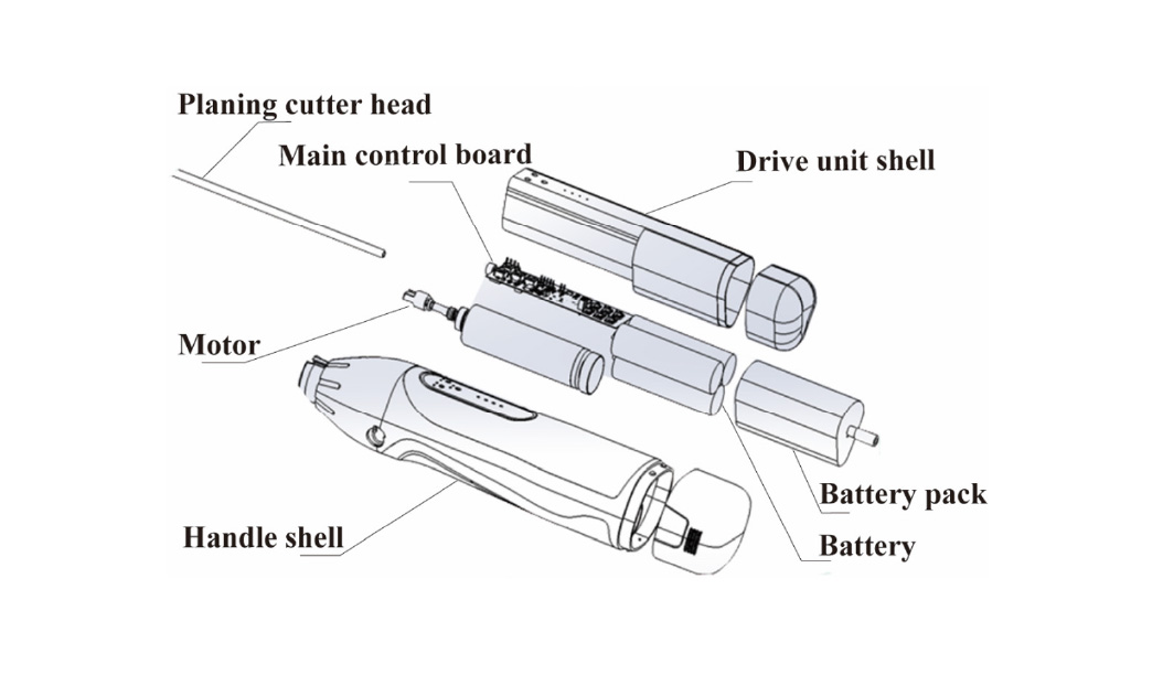
Figure 1. Component units of the portable endoscopic shaving system.
3.2 Performance testing
3.2.1 No-load speed testing
In order to measure the speed range of the product, we connect the rotating parts of the product to the tachometer. After the operator starts the equipment, the change range of the product’s set no-load speed is recorded by adjusting the no-load speed button, and then the speed accuracy is verified. During the test, the operating voltage is set to a rated value and kept within ±2% to ensure voltage stability. The accuracy measurement is carried out at the three positions of the maximum value, the midpoint value, and the minimum value in the speed adjustment range. The actual no-load speed directly reads the tachometer. Each measurement was repeated three times, and the average value was taken as the final result.
Let ns represent the preset no-load speed, nr the measured no-load speed, and Ans the deviation between the preset and actual values. When ns>1,000 rpm, the relative error was calculated as Ans=(nr-ns)/ns×100%; when ns<1,000 rpm, the absolute error was defined as Ans=nr-ns.
3.2.2 No-load reciprocating frequency testing
After device activation, the operator adjusted the no-load reciprocating frequency control to determine the adjustable range and evaluate its accuracy. During the test, the operating voltage was maintained within ±2% of the rated value, while the no-load speed was set at 1,000 rpm. Accuracy measurements were performed at the maximum, midpoint, and minimum frequency settings. The actual reciprocating frequency was directly read from the reciprocating frequency tester. Each measurement point was tested three times, and the average value was calculated. Let vs represent the preset no-load reciprocating frequency, vr the measured no-load reciprocating frequency, and Avs the deviation. When vs≥100 cycles/min, the relative error was calculated as Avs=(vr-vs)/vs×100%; when vs<100 cycles/min, Avs=vr-vs.
3.2.3 Mechanical characteristics testing
A hysteresis dynamometer was used to evaluate the load characteristics of the device. The device was operated at a constant voltage (±2% of the rated value) with a preset speed at 3,000 rpm. Load torque was sequentially applied at 0, 10, 20, 30, and 45 mN·m, followed by gradual increase to the rated load value, while the corresponding rotational speed at each torque level was recorded. Based on these parameters, the overload protection function was tested by applying a load torque to 1.6 times the rated torque. The response of the device was observed to determine whether the overload protection mechanism was activated within 5 seconds to stop the torque output.
3.2.4 Flow rate testing of suction channel
We connected the suction channel of the handle to the electric suction device. After the operator started the device, the voltage stability was controlled within ±2%. Then the aspirator was turned on to adjust the negative pressure to -70 kPa±10 kPa. Then we opened the suction pipe control valve, and placed the planing cutter head in water for suction. The operator recorded the volume (V) and duration (t) of the suction liquid, and calculated the flow rate (L) by the formula L=V/t.
3.2.5 Noise testing
We used a sound level meter to measure the noise level of the equipment. After the product was started, its speed was set to 5000 rpm. Subsequently, the operator recorded noise values in all directions at a distance of 1 meter from the product.
3.2.6 Electromagnetic compatibility testing
This section conducted patient leakage current, casing leakage current, and withstand voltage tests on the product in accordance with the GB 9706.1-2020 [17].
3.2.7 Electrical safety testing
According to the YY 9706.102-2021 standard, we used the measurement receiver, the fixed attenuator, and the 3-meter anechoic chamber to test the electrical safety of the product [18].
3.2.8 Planing cutter head testing
The planing cutter head was connected to the pointer dynamometer, ensuring the test force was applied at the specimen’s mechanical axis or its extension line. We applied stress, observed the force exerted on the cutter head, and recorded the reading.
3.3 Comparison with clinical products
This experiment utilized tissue from animals that had already been sacrificed, rather than live laboratory animals, and therefore did not require animal welfare ethics review. We used fresh pork hooves to evaluate the shaving performance of the device. Fresh pork hooves were placed in deionized water to clean. After that, the surface moisture was removed and the hooves were positioned on the table. The portable device and clinical products were activated, set to the same rotational speed, and used to shave the surface of the pork hooves for 2 minutes. Photographs of the pork hooves were taken, and the area of the shaved regions was statistically measured.
4.1 Composition and structure of the endoscopic shaving system
As a commonly used medical device, the endoscopic shaving system can efficiently remove diseased tissues and alleviate orthopedic-related diseases. The traditional endoscopic shaving system is bulky in structure and has many components. It is mainly composed of a host, a pedal switch, and a handle connected by cables (Figure 2). The traditional structural design makes it difficult to transport and disinfect the equipment, which limits its wider clinical application.
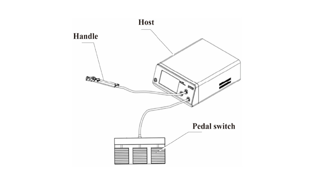
Figure 2. Schematic diagram of conventional endoscopic shaving systems.
In response to this clinical pain point, this work innovatively developed a compact, safe, and portable endoscopic shaving system to replace traditional systems (Figure 3). The portable system can be divided into three parts: drive unit, handle, and planing cutter head. Each component realizes convenient installation, disassembly, and cleaning through the snap-on connection mechanism. The compact design facilitates portability and simplifies intraoperative handling.
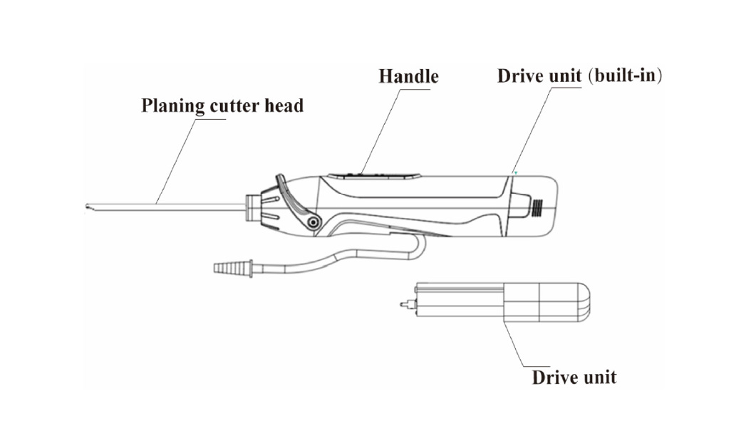
From a structural perspective, the detachable handpiece and shaver blade are the components exposed to the surgical environment, which are designed as disposable units, reducing the risk of bacterial contamination. The detachable handpiece housing incorporates a negative-pressure suction channel and three control buttons, which control the operational state of the shaver blade for effective tissue removal. Simultaneously, the suction channel generates localized negative pressure to facilitate continuous removal of tissue debris during the shaving process.
As the core power module, the drive unit is enclosed within the handpiece to prevent exposure to the external environment. Therefore, there is no need for a sterilization, which improves operational efficiency. In addition, this design eliminates the need for high-temperature-and high-pressure-resistant electronic components, reducing manufacturing costs. Finally, the assembled device can be wirelessly connected to a tablet computer via Bluetooth, enabling functional parameter configuration and real-time operational monitoring.
4.2 Performance testing
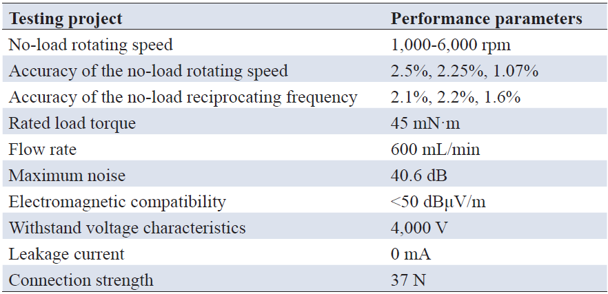
As a critical core module, the drive unit provides mechanical energy required for the endoscopic shaving system. Therefore, systematic performance testing was first conducted on the drive unit. The rotational speed was measured via a tablet interface. The adjustable speed range for both forward and reverse rotation was 1,000-6,000 rpm, while the reciprocating rotational speed range was 1,000-3,500 rpm. Subsequently, the accuracy of preset no-load rotational speeds was evaluated. At preset speeds of 1,000 rpm, 3,000 rpm, and 6,000 rpm, the corresponding deviations (Ans) were 2.5%, 2.25%, and 1.07%, all below 10%, meeting clinical requirements (Figure 4A). Furthermore, the adjustable range of no-load reciprocating frequency was tested, providing discrete frequency settings of 60 times/min (1 s), 67 times/min (0.9 s), 75 times/min (0.8 s), 86 times/min (0.7 s), 100 times/min (0.6 s), 120 times/min (0.5 s), 150 times/min (0.4 s), and 200 times/min (0.3 s). Accuracy evaluation demonstrated that the mean deviations (Ans) at 60, 86, and 200 times/min were 2.1%, 2.2%, and 1.6%, respectively, all within the allowable error threshold (<10%) and meet the clinical application standards (Figure 4B).
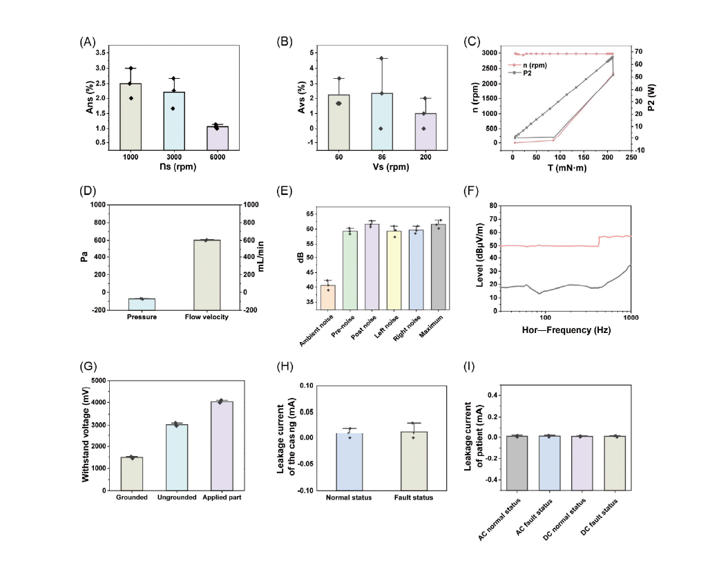
Figure 4. Performance and safety testing of the drive unit and handpiece. (A) Accuracy of preset no-load speed; (B) Accuracy of preset noload reciprocation frequency; (C) Mechanical characteristics testing; (D) Flow rate of the suction channel; (E) Noise levels of the system; (F) Electromagnetic compatibility testing; (G) Withstand voltage testing; (H) Enclosure leakage current testing; (I) Patient leakage current testing.
The team conducted a systematic test of its mechanical properties. At a speed of 3,000 rpm, gradually increasing the load torque from 0 to the rated value of 45 mN·m revealed a noticeable increase in motor power output, while the device maintained a rotational speed of about 3,000 rpm. The product can provide continuous and stable output to meet the needs of practical applications. Due to the existence of the over-protection system, once the load torque exceeds the rated value, the system will close the entire circuit system within 5 s, stop the output, and ensure the safety of users and patients (Figure 4C). The experimental results in this section show that the drive unit exhibits excellent performance indicators in all parameters, and its parameter range design is reasonable, which meets clinical requirements and renders it suitable for further experimentation.
In the handle unit test, the operator can start the planing cutter head by pressing any of the three buttons on the handle to control its forward, backward, and reciprocating rotation motion. When the planing cutter head rotates, pressing any button can immediately stop the power output, indicating that the handle is flexible in operation and reliable in performance. Flow rate testing results also show that the suction channel produces a negative pressure of -70 Pa during operation, and the flow rate can reach 600 mL/min, far exceeding the standard of 400 mL/min, which can effectively remove debris (Figure 4D).
In the noise test, under normal working conditions, the environmental noise generated by the device is 40.6 dB, and the maximum noise is less than 65 dB (Figure 4E). The noise output complies with standards and will not affect patients or operators. Through systematic experimental testing and data analysis, our group confirmed that the handle unit has the advantages of convenient operation, smooth fluid flow, and low noise, and can be applied to subsequent clinical trials.
In the electromagnetic compatibility test, the device can produce stable power output under normal conditions, and the generated signal is less than 50 dBμV/m, which will not cause electromagnetic interference to the surrounding environment and will not interfere with the operation of other surgical instruments (Figure 4F). In addition, the product also shows excellent electrical safety. In the withstand voltage experiment, the equipment can still operate stably under the extreme voltage of 4,000 V, and has strong withstand voltage characteristics (Figure 4G). The leakage current detection results show that the device will not produce leakage current in any state, which fundamentally prevents the occurrence of electrical accidents and ensures the safety of doctors and patients (Figure 4H, 4I).
In order to test the planing cutter head, our team used a pointer dynamometer to measure the connection strength after the final assembly. With the increase of the pulling force, the planing cutter head remains stable and does not fall off, and it can withstand a 37 newton pulling force. After the tension test, the tool head and the handle are perfectly fused, and the operation is normal, and there is no abnormal phenomenon of sticking. This confirms the stability of the system connection and can effectively prevent the occurrence of shedding accidents during use.
Following comprehensive and systematic experimental testing, the parameters of the product’s drive unit, handle, and planing cutter head are summarized in Table 1. The results demonstrate that the product is well-designed and suitable for application in orthopedic surgical scenarios. In addition, the product adopts a wireless structure design, which simplifies the product structure and solves the clinical application shortcomings of traditional instruments, such as difficulty in transportation, infection, and high cost. Consequently, the product has broad clinical application prospects.
4.3 Comparison with clinical products
In this chapter, we compare the clinical firstline equipment with the portable device to discuss whether the device has clinical alternative value (Table 2). There are obvious differences in the appearance and structure between the two. The traditional equipment is mainly composed of the host, the handle, and the pedal switch connected in turn by wires. The portable product adopts a detachable structure design, integrates all modules into a disposable handle, eliminates cable links, simplifies the overall structure of the device, and facilitates the operation and transportation of the device. In the speed adjustment range, the speed range of traditional equipment is 800-8,000 rpm with an error of less than 10%, which can only accept specific speed settings and cannot achieve continuous adjustment. The speed range of this product is 1,000-6,000 rpm with an error of less than 10%, which is lower than that of clinical products, but it can achieve continuous speed adjustment, which can better meet the actual surgical scene.
Table 2. Comparative analysis of the endoscopic shaving systems
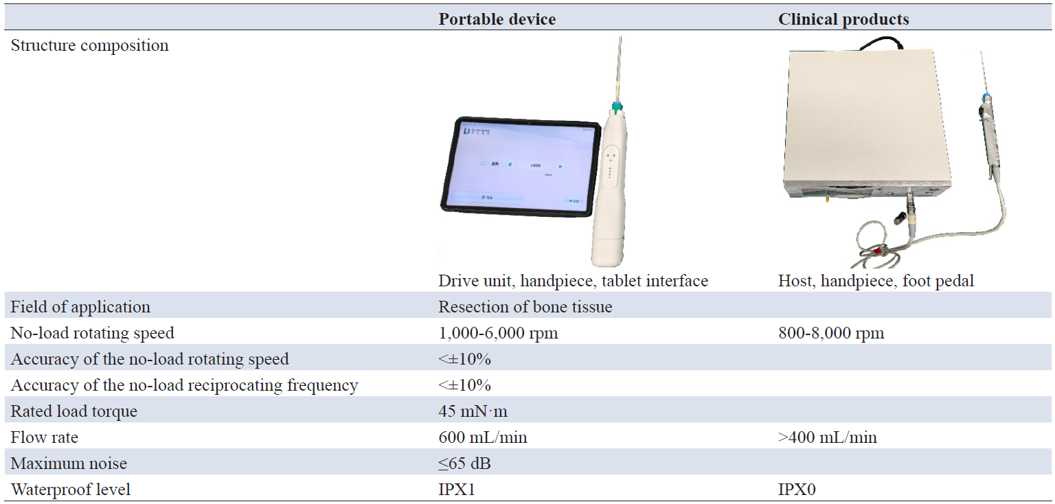
In terms of load characteristics, there are also certain differences between the two products. Comparison reveals that the rated load torque of the traditional product is 116.7 mN·m, while this product’s rated load torque is 45 mN·m, lower than the traditional product but still meeting clinical standards. In the design of the suction pipe, the flow rate of the traditional product can reach more than 400 mL/min, while the flow rate of the product is 600 mL/min. Both of them have the same performance and can quickly remove the tissue debris generated by the operation. At the same time, the noise generated by the product is less than 65 dB in the working state, which is consistent with the clinical product and reduces the interference of noise to patients and operators. In addition, in terms of waterproof performance, the traditional equipment has a waterproof level of IPX0, which cannot resist the interference of tissue fluid on the instrument during the operation, and has certain safety hazards. The product adopts a waterproof process, and the waterproof level reaches IPX1, which avoids the interference of tissue fluid and has high safety.
We used a fresh pig’s hoof as a human tissue simulant to compare the planing performance of the two products. The red box is a portable product planing, and the black box is a clinical product planing position. It can be observed that after continuous planing, both products can produce holes on the pig’s hoof, and the depth morphology of the two products is basically the same (Figure 5A). After that, we used a ruler to measure the void area produced by planing. This product can produce a shaving area of 18 square centimeters, slightly higher than the 16 square centimeters of the clinical product, which can efficiently shave cartilage tissue (Figure 5B).
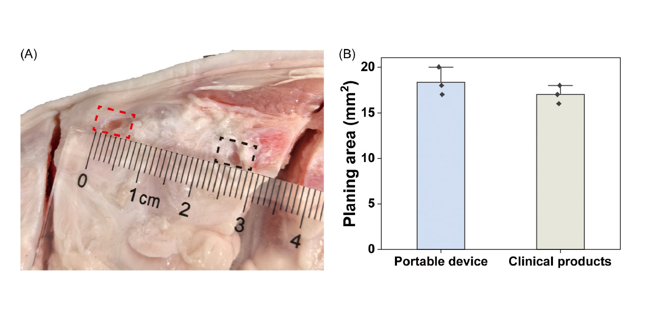
Figure 5. Evaluation of shaving performance. (A) Representative photographs after shaving; the red dashed box indicates the shaving result for the portable device; the black dashed box indicates the shaving result for the clinical product; (B) Quantitative analysis of the shaving area.
Combined with the above data, it can be concluded that the product eliminates the dependence of the device on cables through a unique structural design, realizes wireless operation, facilitates transportation, and reduces the risk of infection. At the same time, the parameters of the product are basically consistent with the clinical first-line equipment, and it has great potential for clinical substitution.
Through the discussion of the above chapters, we demonstrate that this device exhibits excellent planing performance, mechanical properties, and safety, consistent with clinical products. This portable instrument has reached the clinical standard and is very suitable for minimally invasive orthopedic surgery, possessing significant clinical application potential. The detachable structure of the product greatly optimizes the volume of the equipment, reduces the manufacturing and transportation costs of the product, and meets the needs of temporary emergency surgical treatment. In addition, the detachable structure makes the equipment get rid of cable dependence and avoids the problems of mechanical winding and limited operation caused by the cables. At the same time, after the product is disassembled, it can be thoroughly cleaned and fully disinfected to avoid the generation of incomplete cleaning and greatly reduce the risk of bacterial infection.
In addition, orthopedic arthroscopic surgery, as a minimally invasive surgery, often has a small section, and doctors often choose multiple surgeries to perform continuously, which has extremely high requirements for cleaning and disinfection efficiency. Through the detachable design, the product replaces the disposable planing cutter head and the disposable handle shell in time, saves the post-operative disinfection time, improves the operation efficiency, and releases valuable medical resources. In contrast, traditional clinical equipment needs to be disinfected with high temperature and high pressure after surgery, which makes it difficult to clean and disinfect for a long time, affecting the efficiency of surgery. At the same time, the sterilization conditions of high temperature and high pressure have higher requirements for its drive unit, which relies heavily on high-end motor components and increases the cost of equipment manufacturing. Through the detachable design, the product uses a disposable handle to wrap the drive unit inside without additional sterilization, which reduces the dependence on high-end motors and reduces the production cost.
Aiming at the clinical pain points of traditional products, this paper gives the product the advantages of strong portability, stable performance, and low cost by virtue of its exquisite structural design, which not only reduces the complexity of endoscopic surgery but also improves the efficiency of surgery, but also greatly broadens the application scenarios. At the same time, the design also fundamentally solves the problems of bacterial infection in the operation process and improves the safety of the operation. Finally, the product gets rid of the dependence on expensive motors, greatly reduces the cost of treatment, is conducive to the popularization of clinical application, and will benefit more patients in the future.
Aiming at the clinical pain points of traditional endoscopic systems, such as poor transportability, easy infection, and high cost, this paper creatively proposes and designs a portable endoscopic shaving system for the treatment of minimally invasive orthopedic surgery. Through innovative structural design, all modules are integrated into a disposable handle unit, achieving compact size, wireless functionality, and high security. In addition, the performance test data and comparison with clinical products show that the performance of each unit of the product has been basically the same as that of clinical products, all of which have reached the clinical standard, but at the same time, it avoids the problems existing in the practical application of traditional products, has a very high clinical replacement value, and also provides a new research direction for the future research and development of surgical instruments.
Author contributions
Chao Qi was responsible for conducting experiments, collecting relevant data, and performing analysis. Kunyu Chen processed the data, prepared figures, and wrote the manuscript. Jianfei Sun contributed to the project design and proofread the manuscript.
Funding
This research received no external funding.
Data availability
The data that support the findings of this study are available from the corresponding author.
Ethics approval and consent to participate
Not applicable.
Consent for publication
Not applicable.
Competing interests
The authors declare that they have no competing interests.
Acknowledgements
This work was supported by Suzhou Dajiang Medical Technology Co., Ltd.
[1] Shiroshita H, Inomata M, Takiguchi S, Akira S, Kanayama H, Yamaguchi S, et al. Update on endoscopic surgery in Japan: Results of the 16th National Survey of endoscopic surgery by the Japan Society for Endoscopic Surgery. Asian J Endosc Surg. 2024 Apr;17(2):e13285. https://doi.org/10.1111/ases.13285
[2] Mariani PP. The 45 degrees arthroscope: A forgotten scope in knee surgery. Orthop Traumatol Surg Res. 2019 Jun;105(4):691-695. https://doi.org/10.1016/j.otsr.2019.02.003
[3] Wu W, Xie Y, Xue Y, Cui M, Zeng X, Zhang Y, et al. Advantages of 12∘12∘ endoscope in unilateral bipolar endoscopic surgery for lumbar disc herniation. Int J Spine Surg. 2025 Oct 27;19(5):569-577. https://doi.org/10.14444/8771
[4] Ganiev RF, Glazunov VA, Glushkov PS, Levin SV, Shalyukhin KA. Development of new tools for endoscopic surgical operations. J Mach Manuf Reliab. 2023 Jun;52(3):256-261. https://doi.org/10.3103/S1052618823030056
[5] Browning CM, Cloutier R, Rich TC, Leavesley SJ. Endoscopy lifetime systems architecture: Scoping out the past to diagnose the future technology. Systems. 2022 Oct;10(5):189. https://doi.org/10.3390/systems10050189
[6] Ge PS, Lee LS. Advances in endoscopic techniques and technology: Old problems, new frontiers. Dig Dis Sci. 2022 Mar 17;67(5):1429-1430. https://doi.org/10.1007/s10620-022-07435-6
[7] Williams J, Ustin J, Calcei J, Gardiner K, Voos J, Polito P. Wireless vs. wired arthroscopic cameras: A comparative or simulation study of efficiency, cost per case, setup, and takedown. J Orthop Exp Innov. 2022 Dec 12;3(2). https://doi.org/10.60118/001c.38662
[8] Rowinski A, von Schreeb J. Decontamination of surgical instruments for safe wound care surgeries in disasters: What are the options? A scoping review. Prehosp Disaster Med. 2021 Oct;36(5):645-650. https://doi.org/10.1017/S1049023X2100090X
[9] Ofstead CL, Heymann OL, Quick MR, Eiland JE, Wetzler HP. Residual moisture and waterborne pathogens inside flexible endoscopes: Evidence from a multisite study of endoscope drying effectiveness. Am J Infect Control. 2018 Jun;46(6):689-696. https://doi.org/10.1016/j.ajic.2018.03.002
[10] Thornhill G, David M. Endoscope-associated infections: A microbiologist's perspective on current technologies. Tech Gastrointest En. 2019 Oct;21(4):150625. https://doi.org/10.1016/j.tgie.2019.150625
[11] Deb A, Perisetti A, Goyal H, Aloysius MM, Sachdeva S, Dahiya D, et al. Gastrointestinal endoscopy-associated infections: Update on an emerging issue. Dig Dis Sci. 2022 May;67(5):1718-1732. https://doi.org/10.1007/s10620-022-07441-8
[12] Southworth PM. Infections and exposures: Reported incidents associated with unsuccessful decontamination of reusable surgical instruments. J Hosp Infect. 2014 Nov;88(3):127-131. https://doi.org/10.1016/j.jhin.2014.08.007
[13] Deshpande A, Smith GWG, Smith AJ. Biofouling of surgical power tools during routine use. J Hosp Infect. 2015 Jul;90(3):179-185. https://doi.org/10.1016/j.jhin.2015.03.006
[14] Rowan NJ, Kremer T, McDonnell G. A review of Spaulding's classification system for effective cleaning, disinfection and sterilization of reusable medical devices: Viewed through a modern-day lens that will inform and enable future sustainability. Sci Total Environ. 2023 Jun 20;878:162976. https://doi.org/10.1016/j.scitotenv.2023.162976
[15] Choi S, El-Hayek K. Endoscopic equipment- from simple to advanced. Surg Clin North Am. 2020 Dec;100(6):993-1019. https://doi.org/10.1016/j.suc.2020.08.002
[16] Pasquale L, Maurano A, Cengia G, Da Massa Carrara P, Germana B, Graziani MG, et al. Infection prevention in endoscopy practice: Comparative evaluation of re-usable vs single-use endoscopic valves. Infect Prev Pract. 2021 Jun;3(2):100123. https://doi.org/10.1016/j.infipr.2021.100123
[17] Li X, Liu S, Li B, Li J, Yu Z, Zhang Y, et al. Revisions of GB 9706.1-2020 medical electrical equipment revision points and medical device supervision application analysis. China Port Sci Technol. 2023 Apr 10;5(4):42-46. https://doi.org/10.3969/j.issn.1002-4689.2023.04.008
[18] Qin L, Xu K, Wu Y. Rectification scheme for dielectric strength and radiation emission of wearable self-regulating low-frequency therapeutic instrument. Chin J Med Device. 2023 Apr 1;36(7):31-33. https://doi.org/10.3969/j.issn.1002-2376.2023.07.009


 Log In

 On This Page
 CITE
CITE