 Log In
Search




 Open Access
Lize Xiong, Shanghai Fourth People's Hospital Affiliated to Tongji University Translational Research Institute of Brain and Brain-Like Intelligence, School of Medicine, Tongji University, No. 1279 Sanmen Road, Hongkou District, Shanghai 200434, China. E-mail: mzkxlz@126.com. Wei Zhang, Department of Anesthesiology, Pain and Perioperative Medicine, The First Affiliated Hospital of Zhengzhou University, No. 1 Longhu Middle Ring Road, Jinshui District, Zhengzhou 450046, Henan, China. E-mail: zhangw571012@126.com. Jianjun Yang, Department of Anesthesiology and Perioperative Medicine, The First Affiliated Hospital of Nanjing Medical University, No. 300 Guangzhou Road, Gulou District, Nanjing 210029, Jiangsu, China. E-mail: yjyangjj@126.com.
Received September 2, 2025; Accepted September 10, 2025; Published March 31, 2026
Open AccessLize Xiong, Shanghai Fourth People's Hospital Affiliated to Tongji University Translational Research Institute of Brain and Brain-Like Intelligence, School of Medicine, Tongji University, No. 1279 Sanmen Road, Hongkou District, Shanghai 200434, China. E-mail: mzkxlz@126.com. Wei Zhang, Department of Anesthesiology, Pain and Perioperative Medicine, The First Affiliated Hospital of Zhengzhou University, No. 1 Longhu Middle Ring Road, Jinshui District, Zhengzhou 450046, Henan, China. E-mail: zhangw571012@126.com. Jianjun Yang, Department of Anesthesiology and Perioperative Medicine, The First Affiliated Hospital of Nanjing Medical University, No. 300 Guangzhou Road, Gulou District, Nanjing 210029, Jiangsu, China. E-mail: yjyangjj@126.com.
Received September 2, 2025; Accepted September 10, 2025; Published March 31, 2026
With advancements in surgical techniques and increasing complexity of patients' underlying conditions, the management of perioperative bleeding and coagulation disorders has intensified. Clinical practice faces issues such as non-standardized monitoring, inconsistent management protocols for specific patient populations, and unclear thresholds for blood transfusion and hemostatic agent administration. Based on evidence-based principles and integrating the latest evidence since 2020, the task force developed this consensus, titled "Expert Opinion on Perioperative Management of Bleeding and Coagulation in Anesthesiology (2026 Edition)". Focusing on the entire perioperative process, it covered coagulation assessment and monitoring, indications for blood product transfusion, rational use of hemostatic agents, and individualized strategies for special populations, including patients on antithrombotic therapy and those undergoing cardiac, neurosurgical, orthopedic or obstetric surgery. It emphasized preoperative risk screening, intraoperative dynamic monitoring, and post-operative balance between hemostasis and thrombosis prevention. Clinical decision-making should integrate the patient's baseline condition, surgical type, and real-time monitoring results. This opinion aims to provide standardized, multi-disciplinary guidance, optimize management protocols, reduce transfusion-related risks and thrombotic complications, and ultimately enhance perioperative patient safety and improve outcomes.
Perioperative hemorrhage and coagulation abnormalities are major challenges in surgical practice. Uncontrolled bleeding can result in significant blood loss, hemodynamic instability, and a cascade of adverse outcomes, including increased morbidity, prolonged hospitalization, and elevated healthcare costs [1]. On the other hand, excessive correction of coagulation deficiencies may lead to thromboembolic events, posing an equally serious threat to patient safety. The perioperative period, therefore, represents a critical window during which balancing hemostasis and thrombosis is essential [2]. Surgical trauma, anesthesia, and the patient's underlying pathophysiological condition often interact in complex ways, frequently causing coagulation disturbances [3]. The management of perioperative bleeding is further complicated by factors such as pre-existing coagulopathies, the use of anticoagulant or antiplatelet medications, and surgical procedures associated with substantial blood loss [4]. Given these challenges, the need for precise, individualized strategies to manage coagulation and hemorrhage has become increasingly urgent.
Recent advances in diagnostic technologies and therapeutic options have revolutionized perioperative coagulation management. Point-of-care diagnostic tools, such as thromboelastography (TEG) and rotational thromboelastometry (ROTEM), enable real-time dynamic assessments of coagulation status [5]. These innovations facilitate point-of-care monitoring and goal-directed therapy tailored to each patient's coagulation profile. Furthermore, the development of new pharmacological agents, including antifibrinolytics and specific reversal agents for anticoagulants, has broadened the therapeutic options available to anesthesiologists. "Anesthesiologist's consensus on perioperative hemorrhage and coagulation management" was first published in China in 2020 [6]. The 2026 updated version integrates the latest evidence in perioperative coagulation management since 2020, building upon the previous edition. It addresses issues in clinical practice, such as non-standardized monitoring, inconsistent protocols for special populations, and ambiguous blood transfusion thresholds, offering optimization and supplementation to provide diagnostic and therapeutic guidance for the perioperative medicine field both domestically and internationally. This consensus is published in both Chinese and English to facilitate clinical practice exchanges across regions and further enhance perioperative patient safety and outcomes.
2.1 Objectives and scope of the opinion
This opinion aims to establish a standardized, evidence-based framework for the perioperative management of hemorrhage and coagulation disorders. It serves as a comprehensive guide for anesthesiologists and other healthcare professionals involved in perioperative care.
2.2 Target population of the opinion
These recommendations apply to both adult and pediatric surgical patients. Special patient populations include individuals with underlying coagulopathies, those receiving anticoagulant or antiplatelet therapy, obstetric patients, and those undergoing major surgeries, such as cardiothoracic, neurosurgical, and orthopedic procedures.
2.3 Target users of the opinion
The target users are medical personnel involved in the perioperative management of various types of surgeries, including anesthesiologists, surgeons, pharmacists, and nurses.
2.4 Clinical topics
The clinical topics encompass comprehensive strategies for coagulation assessment and monitoring using both traditional laboratory tests and advanced point-of-care diagnostics, the diagnosis and management of coagulation disorders and perioperative bleeding complications, evidence-based protocols for the administration of blood products (including red blood cells [RBCs], platelets, fresh frozen plasma [FFP], and cryoprecipitate), the therapeutic use of hemostatic agents such as antifibrinolytics, procoagulants, and anticoagulant reversal agents, as well as management strategies for mitigating the risk of thrombosis and thromboembolism throughout the perioperative period.
2.5 Opinion development team and process
The consensus panel was carefully selected, comprising 16 experts. A structured three-step Delphi methodology was employed to develop the consensus, conducted between June 2024 and July 2025, with the goal of formulating thorough and practical clinical recommendations. Each panel member independently evaluated the proposed recommendations using a 4-point Likert scale, allowing for a quantitative assessment of the importance of each recommendation. Panel members were also encouraged to propose revisions to enhance clarity and content, ensuring that the recommendations were both practical and accessible.
2.6 Literature search
A comprehensive literature search was conducted to ensure that the recommendations were based on robust evidence. Premier databases such as MEDLINE, Embase, CINAHL, and CENTRAL were searched using key terms, including “perioperative bleeding”, “perioperative coagulopathy”, “hemorrhage management”, “coagulation disorder”, “hemostasis”, “anticoagulant reversal”, “transfusion therapy”, “thromboelastography”, “massive transfusion protocol (MTP)”, and “coagulopathy diagnosis”. In addition to database searches, references familiar to the working group and guidelines from national and international bodies were thoroughly reviewed. The working group predominantly relied on systematic reviews, meta-analyses, and randomized controlled trials to generate evidence-based recommendations. However, it was acknowledged that recommendations concerning laboratory and organizational aspects would largely be based on observational studies. In areas with limited evidence, the working group sought to provide practical, actionable advice by leveraging their combined clinical experience to address knowledge gaps.
Effective perioperative hemorrhage and coagulation management begins with a comprehensive medical history and thorough physical examination. Key aspects of the medical history should include personal and family history of bleeding disorders to identify potential hereditary predispositions, an assessment of chronic liver or kidney dysfunction given their significant impact on hemorrhage and coagulation dynamics, and a thorough review of current medications, as many pharmaceuticals can interfere with the coagulation cascade.
Physical examination should focus on identifying signs of bleeding disorders, including purpura, ecchymosis, and subcutaneous hematomas. Combined insights from medical history and physical examination often offer more value than conventional laboratory tests, such as activated partial thromboplastin time (APTT), international normalized ratio (INR), and platelet count in the preoperative setting. While biochemical immunoassays remain the primary screening tool for preoperative coagulation assessment, genetic testing for hereditary coagulation disorders offers additional safety measures across all perioperative phases. Regarding the vitamin K-dependent coagulation factors (II, VII, IX, and X), which play critical roles in maintaining coagulation homeostasis, clinical attention should be directed toward the supply efficiency of their precursors, especially given the rarity of inactivating mutations [7, 8]. This underscores the importance of a holistic patient assessment as a cornerstone for effective perioperative hemorrhage and coagulation management [9].
Hemorrhage and coagulation monitoring within the perioperative context can be categorized as follows.
3.1 Estimation of blood loss
Quantitative blood loss assessment should employ multiple methods, including examining suction canister contents, evaluating hemostatic gauze saturation, and monitoring surgical drainage tube volumes. These comprehensive evaluations provide crucial data to understand the extent of blood loss and guide subsequent management strategies.
3.2 Monitoring of perfusion and oxygen supply to vital organs
A multimodal monitoring approach is essential, going beyond the simple observation of clinical symptoms and signs. Routine monitoring parameters include blood pressure, heart rate, pulse oximetry, and electrocardiogram recordings. In complex or high-risk cases, additional advanced monitoring techniques may be warranted. These can include: echocardiography to assess cardiac function and perfusion, renal function monitoring via urine output to gauge renal perfusion adequacy, cerebral oxygen saturation monitoring to safeguard cerebral perfusion, arterial blood gas analysis to evaluate gas exchange and acid-base balance, and mixed venous oxygen saturation monitoring to assess tissue oxygen extraction and utilization [10-12]. This comprehensive monitoring enables early detection of perfusion deficits or oxygenation impairments, facilitating prompt interventions to optimize patient outcomes.
3.3 Hemorrhage and coagulation function tests
Hemorrhage and coagulation function testing can be divided into two categories: coagulation cascade evaluation and platelet function analysis. For patients with a documented bleeding history or underlying coagulopathies, preoperative standard laboratory test should include prothrombin time (PT), APTT, INR, fibrinogen (FIB), and D-dimer. These tests help stratify the risk of surgical bleeding and guide preoperative medication optimization. When available, viscoelastic testing should complement conventional assays.
Viscoelastic hemostatic assays (VHA), such as TEG and ROTEM, have become indispensable in modern perioperative care [13]. Integrating VHA into surgical protocols enhances blood product stewardship by establishing evidence-based transfusion thresholds for RBCs, platelets, and plasma during surgery, reducing the overuse of these resources. In settings without VHA access, strict adherence to standard laboratory test with predefined transfusion criteria is essential. During surgery, surgeons must perform real-time visual assessments of the operative field to evaluate coagulation status and identify ongoing surgical bleeding. Notably, VHAs provide superior diagnostic utility in identifying postoperative bleeding causes and guiding anticoagulant therapy.
For patients with suspected platelet dysfunction (e.g., preoperative bleeding history or antiplatelet medication), preoperative platelet count and functional testing are recommended for bleeding risk stratification and medication management. Perioperative platelet monitoring has proven effective in reducing bleeding complications and transfusion requirements in trauma and cardiac surgery patients [2, 14]. While modern techniques such as VHA offer rapid and user-friendly testing, bleeding time measurement is no longer recommended due to its poor reliability from multiple confounding factors.
3.4 Point-of-care testing (POCT)
POCT has become integral to perioperative coagulation management, with several clinically valuable modalities available.
(1) Activated clotting time: Activated clotting time provides rapid bedside coagulation assessment, which is critical during cardiopulmonary bypass (CPB) procedures. Real-time anticoagulation monitoring with activated clotting time helps prevent thrombotic or hemorrhagic events.
(2) TEG and ROTEM: As mentioned earlier, TEG and ROTEM have revolutionized coagulation monitoring by offering dynamic visualization of the entire clotting process, from fibrin formation to clot dissolution. These technologies enable data-driven transfusion decisions and targeted hemostatic interventions during surgeries.
4.1 RBCs
A restrictive strategy for RBC transfusion is recommended in perioperative management. For most patients, maintaining a hemoglobin (Hb) concentration greater than 7 g/dL throughout the perioperative period is generally appropriate. Specifically, for hemodynamically stable adult patients, an Hb threshold of 7 g/dL is suggested as the reference value for considering transfusion. However, if Hb levels drop to 10 g/dL, a comprehensive evaluation of the overall clinical context is necessary. This evaluation should consider factors such as cardiopulmonary compensatory capacity, the patient’s metabolic status, and the presence of active bleeding before deciding on RBC transfusion. For patients undergoing cardiac surgery, a threshold of 7.5 g/dL is recommended, while for those undergoing orthopedic surgery, a target of 8 g/dL is more appropriate [15].
In elderly patients and those with impaired cardiopulmonary function, clinical evaluation should focus on the balance between oxygen supply and demand. The decision to administer RBC transfusions to enhance oxygen-carrying capacity should not be based solely on Hb levels. Instead, it should involve a comprehensive assessment that integrates the patient’s overall physiological condition, comorbidities, and the specific demands of the surgical procedure. This holistic approach ensures judicious use of RBC transfusions, optimizing patient outcomes while minimizing the risks associated with excessive transfusion [16].
The West-China-Liu’s Score provides a framework for determining both the Hb concentration, at which transfusion should be initiated, and the target Hb level after transfusion [17]. The specific methodologies for applying the West-China-Liu’s Score are outlined in Table 1. The West-China-Liu’s Score comprises four components.

Table 1. Huaxi perioperative blood transfusion indication score

Note: *Core body temperature is measured at the nasopharynx, oropharynx, tympanic membrane, rectum, or esophagus. Axillary temperature with an additional 0.5 °C is considered core temperature.
(1) Minimum FiO2 to maintain SpO2≥95%: The minimum FiO2 required to maintain SpO2≥95% serves as a key indicator of pulmonary functional reserve, as attempting to measure the lowest room-air SpO2 in patients with poor lung function is clinically unsafe.
(2) Adrenaline infusion rate: The adrenaline infusion rate is defined as the rate of adrenaline (or an equivalent vasoactive agent) required to maintain adequate cardiac output. It is important to note that the specific agent used may vary according to different institutional protocols.
(3) Core body temperature: Core body temperature reflects the level of total body oxygen consumption, providing insight into the patient’s systemic metabolic demands.
(4) History of angina: A history of angina is a critical factor to consider, as it reflects the heart’s high oxygen demand and its particular sensitivity to imbalances between oxygen supply and consumption. In such patients, the severity of angina is a key clinical consideration.
The West-China-Liu’s Score consists of 6 baseline points, with up to 2 additional points from each of four dimensions, yielding a total score ranging from 6 to 10 (scores are capped at 10). The score correlates with Hb concentration levels, ranging from 6 g/dL to 10 g/dL, to determine the RBC transfusion trigger and target. If the final score is equal to or lower than the current Hb concentration, RBC transfusion is not required. If the final score exceeds the Hb concentration, transfusion is indicated, with the required units of RBCs being twice the difference between the score and the Hb concentration. This calculation assumes that one unit of RBCs is derived from 200 mL of whole blood in China, and transfusion typically raises the Hb level by approximately 0.5 g/dL in most adults.
4.2 Platelets
Platelet transfusion plays a crucial role in perioperative management, particularly for patients with thrombocytopenia or platelet dysfunction accompanied by abnormal bleeding [16]. The decision to transfuse platelets is guided by both numerical thresholds and clinical judgment.
(1) Platelet count ≥100×10⁹/L: Platelet transfusion is generally not indicated when the count exceeds this threshold.
(2) Platelet count <50×10⁹/L: Platelet transfusion is strongly recommended. A count below 50×10⁹/L significantly increases the risk of perioperative bleeding, and prophylactic transfusion can effectively reduce this risk.
(3) Platelet count between 50×10⁹/L and 100×10⁹/L: In this intermediate range, the decision to transfuse requires careful consideration of clinical factors. The presence of spontaneous bleeding or excessive bleeding at the surgical site should be evaluated. If such symptoms are present, platelet transfusion may be necessary to improve hemostatic function.
(4) Intraoperative uncontrolled bleeding: If uncontrollable bleeding occurs during surgery and platelet dysfunction is confirmed, immediate platelet transfusion is essential, regardless of the platelet count. In such critical situations, the priority is to restore effective hemostasis and prevent life-threatening hemorrhage.
4.3 Plasma
FFP is the most commonly used plasma product in the perioperative setting and is essential for correcting coagulation factor deficiencies [16]. The following scenarios outline the primary indications for FFP use.
(1) Abnormal coagulation parameters: FFP is indicated when the PT or APTT exceeds 1.5 times the normal range, or when the INR is greater than 2.0, particularly in the presence of diffuse bleeding at the surgical site. Such abnormalities suggest coagulation factor deficiencies, and active bleeding warrants intervention.
(2) Acute massive bleeding: In cases of significant blood loss, or when large volumes of stored blood or RBCs are transfused (approaching the patient’s total blood volume), FFP supplementation is crucial to prevent or correct dilutional coagulopathy.
(3) Coagulation disorders: FFP is essential for patients with congenital or acquired coagulation dysfunction, whether due to genetic predisposition or underlying medical conditions. Addressing coagulation factor deficits is critical to avoid excessive bleeding.
(4) Reversal of warfarin: In urgent situations requiring reversal of warfarin’s anticoagulant effects, FFP can be administered at a dose of 5-8 mL/kg.
(5) Prophylactic use in high-risk procedures: For patients with abnormal coagulation undergoing invasive procedures or high-risk surgeries, prophylactic FFP administration should be considered to reduce hemorrhage risk.
After FFP infusion, comprehensive clinical evaluation and repeat coagulation tests are necessary. Additional doses may be required based on the results to ensure optimal coagulation and patient safety.
4.4 Cryoprecipitate
Cryoprecipitate is a valuable hematological product rich in essential components, such as factor VIII, FIB, von Willebrand factor (vWF), fibronectin, and factor XIII. In cases of severe hemorrhage with FIB levels below 1.5 g/L, cryoprecipitate administration is strongly recommended [16]. This underscores the critical role of FIB in the coagulation cascade, as its deficiency can significantly impair hemostasis.
5.1 FIB
In cases of significant hemorrhage accompanied by a decline in FIB levels or impaired FIB function, the administration of a FIB concentrate is recommended [18]. FIB therapy should be initiated when the plasma FIB concentration is between 1.5 and 2.0 g/L, and TEG or ROTEM testing indicates FIB dysfunction. The initial infusion dose of FIB concentrate typically ranges from 25-50 mg/kg.
5.2 Coagulation factor XIII (FXIII)
If progressive or diffuse hemorrhage persists despite adequate administration of FIB concentrate, and the patient remains in a hypocoagulable state, this may indicate a significant reduction in FXIII activity. In such cases, when FXIII activity is markedly diminished (<60%), it is recommended to administer FXIII at a dose of 30 IU/kg [19].
5.3 Four-factor prothrombin complex concentrate (PCC)
For patients experiencing severe perioperative hemorrhage while on oral anticoagulant therapy (involving coagulation factors II, VII, IX, and X), the administration of PCC in conjunction with vitamin K is recommended [1]. Additionally, genetic testing for VKORC1 and CYP2C9*3 polymorphisms is recommended.
For patients not receiving oral anticoagulants but presenting with hemorrhage and prolonged coagulation times, PCC should be administered at a dosage of 20-30 IU/kg. However, it is important to note that a prolonged PT/INR alone should not serve as the sole criterion for PCC administration, especially in critically ill patients.
For patients prescribed novel oral anticoagulants, such as dabigatran, reversal of the anticoagulant effect may be needed during emergency surgical procedures, interventional maneuvers, or life-threatening hemorrhagic complications. In these situations, the specific antidote Praxbind is the preferred therapeutic option. PCC may also be considered when the reversal effect of other agents is suboptimal. PCC is recommended for reversing the anticoagulant action of Factor Xa inhibitors in urgent clinical scenarios.
5.4 Recombinant activated factor VII (rFVIIa)
rFVIIa is not recommended for prophylactic use due to its potential to increase the risk of thrombosis. However, when conventional surgical and interventional radiotherapeutic approaches fail to achieve hemostasis, or when comprehensive treatment strategies are ineffective, rFVIIa should be considered. It can also be used to address coagulation disorders associated with hypothermia or acidosis. The recommended dosage is 90-120 μg/kg, which may be administered repeatedly if necessary [1].
5.5 Lysine analogues
(1) Tranexamic acid (TXA): TXA is recommended for the prevention and/or treatment of hemorrhage associated with major surgical procedures or fibrinolysis. The recommended dosage is 20-25 mg/kg, which may be repeated or administered as a continuous intravenous infusion at 1-2 mg/kg/h. Prolonged use may increase the risk of seizures [20].
(2) ε-aminocaproic acid: ε-aminocaproic acid has been shown to reduce intraoperative blood loss and transfusion requirements for blood products in cardiac, hepatic, and orthopedic surgeries through perioperative infusion [1].
5.6 Desmopressin (DDAVP)
Desmopressin, a synthetic analog of arginine vasopressin, increases the plasma levels of coagulation factor VIII and vWF, simultaneously enhancing platelet adhesion [21]. However, current evidence does not conclusively support its efficacy in reducing perioperative bleeding or allogeneic blood transfusion requirements in patients without congenital bleeding disorders. Therefore, its use should be reserved for specific scenarios, such as acquired von Willebrand syndrome. Repeated dosing may diminish its therapeutic effects.
5.7 Calcium supplements
Maintaining normal calcium levels (≥0.9 mmol/L) is essential for optimal coagulation. Clinicians should routinely monitor and supplement calcium as needed during the perioperative period to maintain the integrity of the coagulation system.
6.1 Preoperative management
6.1.1 Screening for bleedin
The risk of perioperative bleeding can be assessed using seven predictive factors: hematoma formation (>2 cm), bleeding history, menorrhagia, surgical history, bleeding after tooth extraction or delivery, and family history of abnormal coagulation [22].
6.1.2 Treatment of preoperative anemia
Preoperative anemia should be managed with erythropoietin and other agents. This condition, associated with an elevated risk of allogeneic transfusion and postoperative complications, requires comprehensive evaluation and timely intervention.
Iron deficiency anemia, the most common form of preoperative anemia, should be treated with iron supplementation. The choice and route of iron therapy should be based on the severity of anemia, the preoperative preparation timeline, and individual variations in iron absorption and tolerance. Two types of therapeutic iron exist: inorganic iron (e.g., ferrous sulfate) and organic iron (e.g., iron dextran and ferrous fumarate), with inorganic formulations generally causing more significant side effects.
Oral iron should be taken after meals to minimize gastrointestinal side effects, which are typically mild and manageable. Iron absorption is inhibited by the concurrent consumption of cereals, dairy products, or tea, and enhanced by the intake of fish, meat, or vitamin C. Therapeutic efficacy is evidenced by rising reticulocyte counts (peaking 5-10 days post-initiation), Hb elevation (apparent after two weeks), and normalization of Hb levels (achieved within approximately two months). Iron therapy should continue for 4-6 months after Hb normalization and only be discontinued when serum ferritin levels stabilize within the normal range.
For patients with oral iron intolerance or malabsorption, intravenous iron supplementation should be considered. Available intravenous formulations, including low-molecular-weight iron dextran, ferric carboxymaltose, iron sucrose, and iron isomaltoside, demonstrate comparable effectiveness. Typical dosing involves 1,000-1,500 mg, administered via slow intravenous infusion once or twice daily. Most patients show Hb improvement within three days, with substantial increases observed after two weeks. Although intravenous iron may trigger allergic reactions, severe anaphylaxis remains exceedingly rare [23].
In megaloblastic anemia caused by folate or vitamin B12 deficiency, treatment should focus on correcting the underlying cause while supplementing folic acid or vitamin B12.
6.2 Intraoperative management
6.2.1 Prevention of perioperative hypothermia
Vigilant prevention of perioperative hypothermia should be maintained through proactive warming measures, aiming to preserve a body temperature >36 °C whenever possible. Temperatures <34 °C may impair platelet function and delay thrombin activation [24].
6.2.2 Management of severe acidosis and anemia
Severe acidosis and anemia should be promptly identified and managed. While pH correction does not immediately reverse acidosis-induced coagulopathy, it requires continuous attention during treatment. Coagulation is significantly compromised at pH <7.10, and reduced hematocrit substantially impairs platelet adhesion and aggregation [25].
6.2.3 Autologous transfusion
Autologous transfusion offers distinct advantages over allogeneic transfusion, including prevention of transfusion reactions, blood-borne infections, and immunosuppression. This method remains the only option for patients lacking compatible donor blood. Salvaged autotransfusion uses blood recovery devices to collect, anticoagulate, wash, and filter blood from surgical sites, body cavities, or postoperative drainage before reinfusion [26]. Strict equipment certification and quality standards must be followed during blood recovery processes. Whenever possible, residual CPB circuit blood should be returned to the patient after the procedure. Coagulation parameters should be closely monitored during large-volume autotransfusion [27].
6.2.4 Development of standardized massive transfusion protocols
6.3 Postoperative management
6.3.1 Causes of postoperative coagulation dysfunction
(1) Coagulation factor depletion and consumption: Surgical trauma and physiological hemostatic responses may lead to the consumption of coagulation factors, thereby affecting coagulation function.
(2) Hemodilution: Aggressive fluid resuscitation often results in hemodilution, reducing the concentration of coagulation factors in the circulation.
(3) Hypothermia: Inadequate maintenance of body temperature (<34 °C) can inhibit thrombin activity and platelet aggregation.
(4) Acidosis: Acidosis caused by tissue ischemia, shock, or respiratory failure can impair coagulation factor activity and platelet function.
(5) Pharmacological agents: Medications such as aspirin (irreversibly inhibiting platelet function), clopidogrel (interfering with platelet activation), and low molecular weight heparin (LMWH) (for thromboprophylaxis) may alter coagulation status. Attention should be paid to their cumulative effects, and monitoring should be intensified.
6.3.2 Clinical manifestations of postoperative coagulation dysfunction
The primary manifestation of postoperative coagulation dysfunction is postoperative hemorrhage. In the immediate postoperative period, the characteristic sign of fresh blood permeating through the incision dressings may be observed, along with an increased flow rate in the surgical drainage tube. Patients may exhibit physiological responses such as tachycardia, hypotension, or require vasoactive support to maintain hemodynamic stability. Additional signs may include local petechiae, ecchymosis in surrounding tissues, spontaneous epistaxis, gingival hemorrhage, occult gastrointestinal bleeding, or hematuria [29].
Accurately determining the underlying cause of postoperative hemorrhage is essential for prevention. The surgical procedure itself should be thoroughly assessed, and sophisticated diagnostic techniques (e.g., computed tomography) may be required. Immediate surgical intervention, either through direct hemostasis or interventional embolization, should be initiated without delay, even if overt coagulopathy has not been diagnosed. Swift control of bleeding can prevent further clinical deterioration.
Surgically induced postoperative hemorrhage may develop into more severe coagulopathy if not promptly addressed. However, in many cases, bleeding can be controlled by correcting the surgical issue and providing appropriate symptomatic treatment. Postoperative hemorrhage due to an underlying coagulation disorder requires comprehensive therapeutic strategies, as surgical reintervention alone is insufficient.
6.3.3 Prevention of postoperative venous thromboembolic complications
Venous thromboembolic complications, including deep venous thrombosis and pulmonary embolism, are potentially life-threatening. Understanding the associated risk factors is crucial for implementing effective prophylactic strategies [30].
(1) High-risk factors: hip and knee arthroplasty, complex general surgeries, pelvic or femoral fractures, severe multi-system trauma, and spinal cord damage, all of which result in prolonged immobility, compromised endothelial integrity, and hypercoagulable states.
(2) Moderate risk factors: arthroscopic knee procedures, pregnancy and the postpartum phase (characterized by hormonal fluctuations and venous stasis), malignant tumor (malignancy-related hypercoagulability), chemotherapy (which can affect coagulation function), central venous access devices, and a history of venous thromboembolic events.
(3) Low-risk factors: laparoscopic surgery, obesity, age over 50 years, bed rest exceeding 3 days, prolonged sitting, and lower extremity varicose veins.
Drugs involved in prevention and treatment [31-35]:
(1) Heparin: A fast-acting anticoagulant that can be rapidly neutralized by protamine when reversal of the anticoagulant effect is required, but may predispose patients to bleeding tendencies and thrombocytopenia; coagulation function and platelet counts should be monitored during postoperative use.
(2) LMWH: A commonly used postoperative anticoagulant widely employed in orthopedic and vascular surgery, which can be administered as early as 6 hours after surgery to significantly reduce the incidence of postoperative deep vein thrombosis without increasing bleeding risk. It offers more rapid absorption subcutaneously, higher bioavailability, and lower risks of bleeding and thrombocytopenia compared to unfractionated heparin, but lacks a specific reversal agent.
(3) Thrombolytics: Agents such as streptokinase and urokinase primarily used for emergency management of thromboembolic complications, particularly pulmonary embolism, but associated with a high risk of bleeding complications requiring cautious use.
7.1 Patients on antithrombotic therapy
An increasing number of patients require perioperative antithrombotic therapy for thrombosis prevention. These patients often have concomitant cardiovascular diseases (such as atrial fibrillation, coronary heart disease, or post-valve replacement surgery) or a history of venous thromboembolism (VTE). Although antithrombotic agents effectively reduce thrombotic risk, they disrupt the body’s normal hemostatic balance, significantly increasing the risk of perioperative bleeding. This challenge is further compounded by the dual effects of surgical trauma, which induces both coagulation activation and hyperfibrinolysis, making the paradox between bleeding and thrombosis even more complex.
7.1.1 Commonly used antithrombotic agents in clinical practice
7.1.1.1 Antithrombin drugs
(1) Unfractionated heparin: In cases of severe heparin-related bleeding, protamine infusion is required for reversal.
(2) LMWH: Its half-life is 3-4 hours, and protamine can only partially reverse its anticoagulant effect.
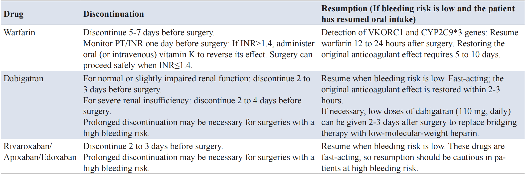
(3) Warfarin: As a vitamin K antagonist (VKA) with a half-life of 2-4 days, its anticoagulant effect takes 5-7 days to fully dissipate after discontinuation. INR is the primary indicator for monitoring its anticoagulant effect, and most surgeries require an INR≤1.4.
(4) Direct oral anticoagulants (DOACs): Classified into direct thrombin (factor IIa) inhibitors and direct factor Xa inhibitors based on their mechanism of action. For procedures with low bleeding risk, DOACs should be discontinued 1-2 days preoperatively. However, for moderate-to-high bleeding risk procedures with a creatinine clearance >50 ml/min, rivaroxaban, apixaban, and edoxaban should be discontinued 3 days prior to surgery, with no requirement for bridging anticoagulation [36, 37]. Specific management strategies should reference the 2024 AHA/ACC/ACS/ASNC/HRS/SCA/SCCT/SCMR/SVM Guideline for Perioperative Cardiovascular Management for Noncardiac Surgery [37].
7.1.1.2 Antiplatelet drugs
(1) Aspirin: Cyclooxygenase inhibitor. Continuation of aspirin therapy is generally acceptable during the perioperative period for most surgical procedures. If preoperative discontinuation is necessary, a 5-7 day cessation period is recommended. Platelet transfusion can be considered for managing aspirin-associated bleeding complications.
(2) Clopidogrel: Adenosine diphosphate receptor inhibitor. Discontinuation of clopidogrel 5-7 days before surgery is critical for patients at elevated bleeding risk. In cases of intraope rative or postoperative clopidogrel-related bleeding, platelet transfusion should be considered as a management strategy.
7.1.1.3 Fibrinolytic drugs
Streptokinase: An exogenous activator of the fibrinolytic system with thrombolytic effects. Treatment depends on selective supplementation of platelets and plasma proteins. Most patients require cryoprecipitate transfusion to replenish FIB, along with platelet transfusion to correct platelet dysfunction.
7.1.2 Perioperative management of antithrombotic drugs
The optimal timing for perioperative discontinuation of antithrombotic drugs remains insufficiently supported by large-scale clinical studies. Current practice relies on empirically derived protocols for medication cessation and reintroduction (see Table 2) [32-35]. Laboratory monitoring plays a critical role in guiding the precise management of specific agents, enabling clinicians to assess coagulation status and optimize therapeutic decision-making.
7.1.2.1 Bridging therapy
Bridging therapy involves the administration of short-acting anticoagulants during the cessation of long-acting anticoagulants. This approach is typically indicated for patients on antithrombotic medications in specific circumstances, including a history of cerebral embolism or systemic embolic events within the past three months, after mechanical mitral or aortic valve replacement (particularly aortic valve replacement with additional stroke risk factors), in patients with atrial fibrillation and high stroke risk, in those with a history of VTE in the past three months, in cases with previous long-term use of antithrombotic drugs where thromboembolic events occurred during the withdrawal period, and during a specific interval after coronary stent implantation.
For patients undergoing minimally invasive procedures with negligible bleeding risks, such as cataract surgery, minor dental procedures, and superficial skin interventions, bridging therapy is generally unnecessary. In patients with an elevated bleeding risk, the initiation of bridging anticoagulation requires careful evaluation, particularly during the postoperative period [38].
The commonly used drugs in bridging therapy are heparin and LMWH. LMWH is more frequently used due to its ease of administration and reduced need for frequent coagulation monitoring. Bridging therapy can be implemented preoperatively, postoperatively, or during both phases. For patients receiving LMWH bridging prior to elective, high-bleeding-risk surgery, it is recommended to administer half the total daily dose of LMWH the day before surgery, rather than the full dosage.
For patients requiring interruption of DOACs for elective surgery, perioperative heparin bridging should be avoided. Resuming DOACs more than 24 hours after surgery is preferable to resuming within 24 hours [39].
For patients undergoing VKA therapy who need VKA interruption for elective surgery, heparin bridging should be avoided in the following cases: atrial fibrillation, mechanical heart valve, VKA therapy whose sole indication is VTE, and colonoscopy with anticipated polypectomy. For patients undergoing pacemaker or implantable cardioverter-defibrillator implantation while on VKA therapy, continuation of VKA therapy is recommended, avoiding interruption with heparin bridging [40].
Conventional bridging therapy is generally unnecessary for most patients with atrial fibrillation. However, for those at high risk of thromboembolic events (e.g., CHA2DS2-VASc score ≥6), bridging therapy should be considered preoperatively and postoperatively. Bridging therapy begins with the discontinuation of the original antithrombotic drug as its effect wanes. It continues until adequate hemostasis is achieved post-surgery, and the original therapy is resumed, restoring its efficacy to pre-discontinuation levels [32].
For patients with prior VTE, if a procedure is required within one month of the acute episode, bridging therapy should be implemented preoperatively and postoperatively. If the episode occurred more than one month prior, bridging therapy is typically only needed after surgery [41, 42].
7.1.2.2 The bleeding risk in neuraxial anesthesia
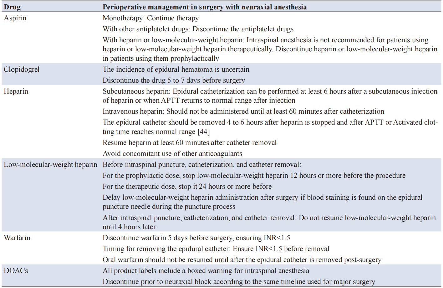
Antithrombotic agents have variable effects on bleeding during neuraxial anesthesia. Given the potential severity of bleeding complications within the spinal canal, such as epidural hematoma, the decision to proceed with neuraxial anesthesia must be made with great caution, guided by a thorough patient evaluation. Neuraxial anesthesia should only be performed by experienced anesthesiologists with precision. Postoperative monitoring of peripheral nerve function is crucial, and any associated complications must be promptly identified and addressed (see Table 3) [43, 44].
7.2 Patients undergoing cardiovascular surgery
7.2.1 Preoperative preparation
Patients receiving antithrombotic therapy should strictly follow the contents above for comprehensive coagulation status evaluation and risk mitigation. Patients with chronic right heart failure and concomitant hepatic dysfunction (impaired coagulation factor synthesis) require preoperative optimization through multimodal medical management, including hepatic support and nutritional supplementation. For urgent surgeries in antiplatelet/anticoagulant-treated patients, immediate availability of allogeneic platelet reserves is essential to counteract medication-induced hemorrhagic risks.
7.2.2 Drugs to reduce bleeding
7.2.2.1 Prophylactic use of antifibrinolytic agents
Synthetic agents such as TXA, ε-aminocaproic acid, and aminomethylbenzoic acid share similar mechanisms and effectively reduce perioperative blood loss, transfusion requirements, and reoperation rates in cardiac surgery. TXA is particularly potent (approximately 10 times more effective than ε-aminocaproic acid), significantly decreasing RBC transfusion needs and the incidence of re-thoracotomy.
For low-risk bleeding surgeries (e.g., simple valvuloplasty, valve replacement, and coronary artery bypass grafting), a loading dose of 10 mg/kg TXA followed by a maintenance dose of 1-2 mg/kg/h is advised.
For high-risk bleeding surgeries, a loading dose of 30 mg/kg TXA and a maintenance dose of 16 mg/kg/h are recommended. Studies suggest that a total dose exceeding 50 mg/kg may be associated with increased postoperative seizures [45]. Therefore, it is not advisable to exceed a total dose of 50 mg/kg in non-high-risk bleeding patients.
It is crucial to use antifibrinolytic drugs prophylactically in patients undergoing CPB. Regardless of the selected dosage regimen, achieving an effective blood drug concentration prior to CPB initiation is essential. For TXA, a minimum intravenous dose of 10 mg/kg should be administered before CPB to maintain adequate blood levels during bypass, with discontinuation of the infusion after CPB [14, 46]. Research indicates that high-dose TXA infusion reduces the proportion of patients requiring RBC transfusions in cardiac surgeries with CPB. High-dose TXA infusion also leads to less postoperative blood loss in patients with acute type A aortic dissection undergoing surgical repair. Furthermore, TXA is associated with a lower risk of bleeding in patients undergoing coronary artery surgery, irrespective of the use of CPB (on-pump vs. off-pump) or the concomitant performance of valve replacement or other cardiac procedures [46]. However, TXA is associated with an increased risk of postoperative seizures [47].
7.2.2.2 DDAVP
DDAVP is the only pharmaceutical intervention effective in managing hemorrhage caused by abnormal platelet function after CPB-assisted cardiac surgery. Indications include patients undergoing coronary artery bypass grafting who received antiplatelet agents within seven days preoperatively or required CPB durations exceeding 140 minutes. The recommended regimen is 0.3 μg/kg intravenously, capped at 15 μg for patients weighing <100 kg. Pharmacodynamic studies show that therapeutic effects begin within 1 hour of administration and persist for approximately 6 hours. Optimal timing involves administering DDAVP about 1 hour before CPB weaning, typically during the rewarming phase. In non-CPB surgery, DDAVP should be reconstituted in 100 mL of normal saline and administered via slow intravenous infusion over 15-30 minutes. During CPB surgery, medications should be administered via slow intravenous infusion to avoid hypotension. Repeated administration may reduce efficacy, as DDAVP stimulates rapid synthesis of biologically active coagulation factors from vWF and factor VIII precursors in the body. It is ineffective when administered at the commencement or before surgery [48].
7.2.2.3 FIB concentrate
This agent plays a crucial role in replenishing FIB levels, particularly when FIB depletion contributes to abnormal hemorrhage. Timely administration can restore normal hemostasis and mitigate further bleeding complications.
7.2.2.4 PCC
PCC provides a concentrated formulation of prothrombin and other coagulation factors, effectively managing perioperative coagulation factor deficiencies in cardiovascular surgery. This treatment restores the coagulation cascade balance and reduces the risk of hemorrhage.
7.2.2.5 rFVIIa
It is used for salvage treatment of refractory abnormal bleeding after CPB surgery [49]. A low-dose rFVIIa regimen (20-40 μg/kg) should be administered alongside FIB, FFP, and platelet supplementation. This combined approach provides hemostatic effects, achieving hemostasis while reducing the risk of thromboembolic complications.
7.2.3 Other coagulation management measures
7.2.3.1 Heparin anticoagulation and protamine neutralization
During CPB, maintaining adequate heparin anticoagulation prevents excessive clotting. However, protamine exhibits platelet-inhibitory and anticoagulant properties, so its administration should be carefully controlled to avoid overdosage. The initial protamine dose for heparin neutralization should be calculated as 1.0:0.5 of the total heparin amount present in the body (including that used during CPB). For example, if the total heparin dosage is 40,000 U, the initial protamine dose should be 200 mg. After initial neutralization, intermittent protamine supplementation or continuous infusion via a pump is necessary to achieve an approximate 1:1 ratio of total protamine to heparin by the end of the surgical procedure. From the initiation of protamine administration until 6 hours postoperatively, the residual effects of heparin should be regularly assessed, with protamine administered as needed based on the clinical situation [37, 39].
7.2.3.2 POCT
TEG and ROTEM are critical diagnostic tools for patients with abnormal bleeding manifestations or suspected coagulation disorders. These techniques help identify the etiological factors underlying bleeding, guide therapeutic interventions, reduce the volume of allogeneic blood transfusions during the perioperative phase, and improve patient prognosis [50].
7.2.3.3 Autologous plateletpheresis
Preoperative autologous platelet separation technology allows for the collection of autologous platelets and RBCs using a blood separation apparatus. RBCs are re-infused based on the patient’s Hb concentration, while platelet-rich plasma is predominantly re-administered to the patient following heparin neutralization [51].
7.3 Patients undergoing neurosurgical procedures
7.3.1 RBC transfusion
Blood conservation strategies in neurosurgery encompass various components, including indications for RBC transfusion, correction of coagulopathies, acute normovolemic hemodilution, autologous blood salvage, use of antifibrinolytic agents, and avoidance of non-steroidal anti-inflammatory drugs.
Traumatic brain injury (TBI), intracranial neoplasms, cerebral hemorrhage, and other conditions can decrease intracranial compliance, disrupting cerebral blood flow regulation. Cerebral blood flow, Hb concentration, blood oxygen content, and blood viscosity are key parameters requiring meticulous monitoring during perioperative management of neurosurgical procedures.
When Hb concentration declines, normal brain tissue compensates by dilating cerebrovascular vessels and increasing cerebral blood flow to maintain stable oxygen supply. However, this compensatory mechanism is limited. When Hb concentration drops to 5-6 g/dL, cerebrovascular dilation reaches its maximum. Further reductions in Hb concentration lead to a decrease in cerebral oxygen supply, resulting in cerebral anoxia and related symptoms [52].
Multiple factors, including TBI, neurosurgical intervention, cerebral hemorrhage, and reduced cardiac output, can impair the brain’s compensatory response to declining Hb concentrations. Therefore, it is not feasible to define a universal compensatory capacity for neurosurgical patients. The indications for RBC transfusion should be based on the pathophysiological changes associated with each neurosurgical disorder and the individual patient’s clinical condition. Recent transfusion strategies for patients with brain injury have become a focal point in neurocritical care and neurosurgical anesthesia, requiring a balance between cerebral oxygen supply and transfusion-related risks. Three randomized controlled trials published in 2024—TRAIN, HEMOTION, and SAHARA—examined liberal versus restrictive transfusion strategies for various brain injury subtypes, offering guidelines tailored to specific contexts [53-55].
Current evidence supports personalized strategies that consider brain injury phenotypes, pathophysiological phases, and real-time monitoring metrics, such as cerebral oxygen saturation or jugular venous oxygen saturation. These approaches aim to optimize neuroprotection while minimizing complications, such as transfusion-related acute lung injury or immunomodulatory effects that could worsen brain inflammation.
(1) Subarachnoid hemorrhage
Anemia affects 40% to 50% of patients with subarachnoid hemorrhage. Research shows that an Hb concentration below 9 g/dL is an independent risk factor for brain tissue damage in these patients [56]. Maintaining a postoperative Hb level of 11-12 g/dL may reduce the incidence of symptomatic cerebral vasospasm after surgery. Subarachnoid hemorrhage patients often exhibit significantly reduced intracranial compliance, and aggressive RBC transfusion strategies have been linked to adverse outcomes. The transfusion of aged RBCs may also increase the incidence of unfavorable consequences in these patients [57].
The HEMOTION trial, focused on acute ischemic stroke and intracerebral hemorrhage, found no significant difference in 6-month neurological outcomes between liberal (<10 g/dL) and restrictive (<7 g/dL) transfusion thresholds [53]. Although no overall benefit was observed, survivors in the liberal group showed modest improvements in functional independence, highlighting potential subgroup effects that warrant further exploration.
(2) TBI
Due to the heterogeneous nature of injury mechanisms, components, and severities, TBI patients exhibit varying degrees of attenuation in the brain’s ability to compensate for anemia. Research indicates that TBI patients with anemia experience poorer recovery of neurological function at 3 and 6 months post-injury [52]. Moreover, the mortality rate is significantly higher in patients with Hb concentrations <9 g/dL. TBI patients who receive allogeneic RBC transfusions are also predisposed to a higher incidence of infection, mortality, and postoperative neurological dysfunction.
For TBI patients with Hb concentrations ≥10 g/dL, allogeneic RBC transfusion is not recommended. For patients with Hb concentrations <7 g/dL, RBC transfusion should be administered. If the Hb concentration falls between 7 and 10 g/dL, RBC transfusion should be considered based on cardiac and pulmonary reserve capacity. Additionally, transfusion should be considered when the brain tissue partial pressure of oxygen decreases below 20 mmHg (1 mmHg=0.133 kPa) or when cerebral oxygen saturation drops below 60% [58, 59].
The TRAIN trial, focused on TBI, compared a liberal transfusion threshold (Hb <9 g/dL) with a restrictive threshold (<7 g/dL). The liberal strategy resulted in a 10% reduction in the risk of unfavorable neurological outcomes at 6 months (62.6% vs. 72.6%) and a 35% decrease in cerebral ischemic events [55]. These findings suggest that maintaining higher Hb levels may mitigate secondary brain injury by improving cerebral perfusion, particularly in TBI patients with disrupted oxygen metabolism. The SAHARA trial, which examined aneurysmal subarachnoid hemorrhage, did not show superiority of the liberal transfusion strategy (for Hb<10 g/dL) over restrictive strategies (Hb<7 g/dL) regarding 12-month modified Rankin Scale scores. This emphasizes the need for caution in this group, as hypervolemia may worsen cerebral vasospasm or edema [54].
These findings underscore the complexity of transfusion decision-making in brain injury. The optimal transfusion thresholds depend on injury type, pathological stage, and individualized monitoring. For mixed or moderate-severe TBI, the TRAIN trial supports a 9 g/dL threshold to balance oxygen delivery and reduce ischemic risk. However, for aneurysmal subarachnoid hemorrhage, the neutral results of SAHARA caution against over-transfusion, as the risk of adverse hemodynamic effects may outweigh potential benefits.
7.3.2 Platelet transfusion
7.3.3 Correction of coagulation disorder
Patients with acute craniocerebral trauma frequently develop acute traumatic coagulopathy, defined by INR>1.3, PTT>38 seconds, or platelets <100×109/L within 7 days of injury. Acute traumatic coagulopathy occurs in 20.6% of patients and is associated with higher rates of shock, hypothermia/hyperthermia, and coagulation derangement. These patients require more blood products, which correlates with increased in-hospital mortality, longer hospital stays, higher incidences of deep venous thrombosis, seizures, and long-term disability. Limited evidence exists regarding the benefits of FFP and PCC for improving coagulation function and prognosis in these patients [61].
TXA can reduce blood loss and the proportion of patients requiring allogeneic transfusion in neurosurgical procedures; however, it may increase the incidence of perioperative seizures [62-64]. Currently, there is insufficient evidence to determine the optimal dosage of TXA for perioperative management in neurosurgery [65].
The administration of non-steroidal anti-inflammatory drugs during elective craniotomy may increase the risk of postoperative intracranial hemorrhage. Thus, non-steroidal anti-inflammatory drugs should be used cautiously during the perioperative period of brain parenchyma surgery in neurosurgery [66].
7.3.4 Autologous blood transfusion
Autologous blood transfusion includes preoperative autologous blood donation, intraoperative cell salvage, and acute normovolemic hemodilution. The indications and contraindications for autologous blood transfusion in neurosurgery are as follows.
Indications: cerebrovascular surgery with anticipated intraoperative blood loss reaching 20% or more of the patient’s blood volume, hemispherectomy, surgery for craniosynostosis, and surgery for closed craniocerebral injury.
Contraindications: surgeries with a contaminated operative field (such as transoral and transnasal approaches, evacuation of intracranial or intraspinal abscesses/other infectious foci, and open craniocerebral trauma surgery); surgeries for malignant tumors. Autotransfusion should be used with caution in patients with meningioma, as there have been multiple case reports of ectopic implantation and metastasis of meningioma cells via blood passage through the pulmonary circulation. For meningiomas located near the cerebral venous sinuses, the risk of tumor metastasis associated with autotransfusion must be weighed against the life-threatening risk of massive hemorrhage. Autotransfusion should only be considered in life-threatening emergencies with massive bleeding, when allogeneic blood is unavailable [67].
7.4 Patients undergoing orthopedic surgery
Blood loss varies considerably across orthopedic procedures. Surgeries involving pelvic fractures, femoral shaft fractures, total knee arthroplasty, and spinal interventions are particularly associated with substantial perioperative hemorrhage. Elective orthopedic surgery patients should follow standard perioperative blood management principles, including correcting preoperative anemia, implementing acute normovolemic hemodilution, utilizing intraoperative autologous blood salvage, maintaining normothermia, applying controlled hypotension, optimizing surgical techniques, and managing postoperative anemia. Restrictive transfusion protocols are recommended to minimize allogeneic blood product exposure.
7.4.1 Trauma orthopedic patients
7.4.2 Antifibrinolytic and anticoagulant treatment for orthopedic surgery patients
Orthopedic procedures often involve substantial blood loss, including both visible intraoperative hemorrhage and concealed blood loss due to surgery-induced fibrinolysis, which should not be overlooked [68-70]. Given the high risk of VTE associated with these surgeries, anticoagulation therapy becomes necessary—a process that requires a delicate balance between antifibrinolytic and anticoagulant treatments [13].
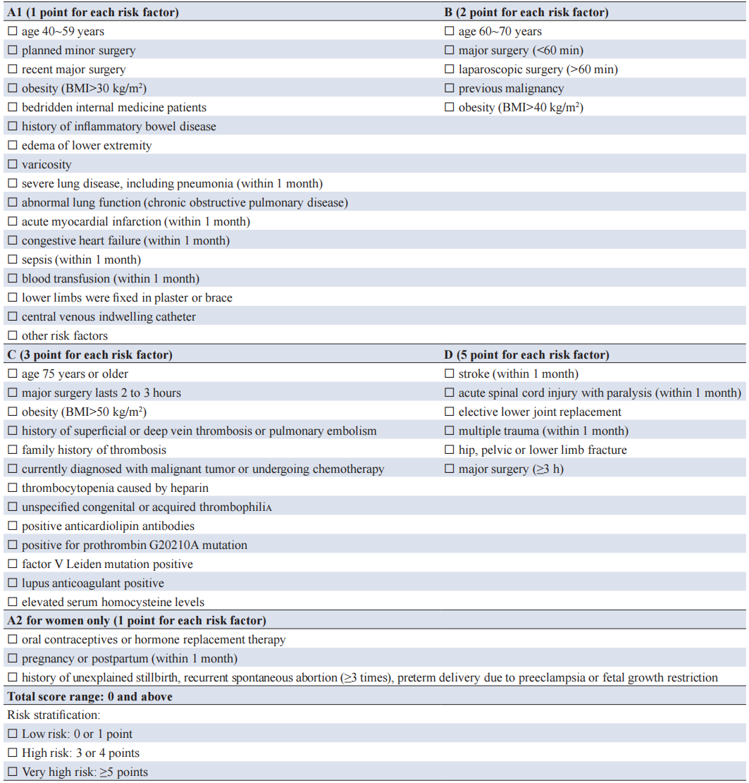
TXA should be administered perioperatively to patients undergoing hip and knee replacement surgery, either intravenously or topically. Similarly, intravenous TXA administration is advised perioperatively for patients undergoing spinal surgery [13]. In hip and knee replacement surgeries, anticoagulants should be administered 6-12 hours after bleeding cessation. If significant bleeding persists 12 hours postoperatively, the initiation of anticoagulation may be postponed [61, 66]. Before spinal surgery, thrombosis risk should be assessed using the Caprini score (Table 4) [67]. For patients receiving TXA during the perioperative period, the following recommendations regarding anticoagulant administration are proposed: For high-risk patients, anticoagulants should be initiated 12 hours after surgery once bleeding has ceased; For intermediate-risk patients, anticoagulants should be initiated 12 to 24 hours after surgery once bleeding has ceased; For low-risk patients, only basic preventive measures and physical methods are recommended. In high-risk bleeding patients, anticoagulation may be delayed until 24 hours post-surgery or potentially withheld entirely [61, 66]. Generally, anticoagulant medications should be administered for 10-14 days. However, in patients with a high VTE risk post-surgery, the duration may be extended to 15-35 days. When abnormal coagulation or hemorrhagic episodes occur, a comprehensive evaluation of bleeding and thrombosis risks is essential, with medication dosages adjusted or discontinued as necessary.
7.5 Patients undergoing obstetric surgery
7.5.1 Assessment of postpartum hemorrhage (PPH)
PPH is defined as blood loss exceeding 500 mL within 24 hours after vaginal delivery or surpassing 1,000 mL after a cesarean section. However, accurately quantifying blood loss during the perinatal period remains challenging in clinical practice. A comprehensive systematic review suggests that improving the precision of blood loss assessment alone may not be sufficient to prevent or manage PPH effectively [71]. Nevertheless, research has shown that implementing standardized bleeding management protocols and promptly preparing blood products can reduce the need for blood transfusions [72].
7.5.2 Coagulation monitoring
7.5.3 Component transfusion
The current consensus for RBC transfusion in obstetric patients is to maintain an Hb level above 8 g/dL, with a recommended platelet count of at least 75×10⁹/L. Plasma FIB levels should be maintained at relatively high levels, with a threshold exceeding 2 g/L. In the context of MTP, a 1:1:1 ratio of erythrocytes, plasma, and platelets is typically employed. However, for patients with PPH, this ratio may not always be applicable, as there is limited research supporting its efficacy in this population [72].
7.5.4 Intraoperative cell salvage
Recent studies and clinical evidence have demonstrated the safety of intraoperative cell salvage in obstetric patients. Indications include limited blood supply or difficulty in cross-matching; cesarean section with anticipated blood loss exceeding 20% of total blood volume; and patients who decline allogeneic blood transfusions due to religious beliefs or personal preferences. During the procedure, two separate suction devices should be used—one exclusively for amniotic fluid collection. The salvaged blood must be processed through a certified filter before reinfusion. For Rh-negative pregnant women, intraoperative cell salvage is recommended when anti-D immunoglobulin is available [73].
7.5.5 Interventional therapy
7.5.6 Amniotic fluid embolism with disseminated intravascular coagulation (DIC)
Classic amniotic fluid embolism manifests as sudden cardiopulmonary collapse, pulmonary hypertension, and coagulation dysfunction during labor or immediately after delivery. While maintaining respiratory and circulatory function, anesthesiologists should assess coagulation status as early as possible and actively correct coagulation disorders. For patients complicated with DIC, RBCs and coagulation factors should be promptly supplemented according to MTP. In cases of hyperfibrinolysis (particularly hypofibrinogenemia), priority should be given to FIB supplementation, along with early administration of TXA for antifibrinolytic therapy. The hypercoagulable phase of DIC is difficult to identify clinically; therefore, heparin therapy is not routinely recommended unless there is clear evidence of a hypercoagulable state [75].
7.5.7 TXA
Balanced management of perioperative bleeding and coagulopathy is one of the core pillars for ensuring the safety and improving the prognosis of surgical patients. Its complexity has become increasingly prominent alongside the advancement of surgical techniques, the diversification of patients’ underlying comorbidities, and the widespread clinical application of antithrombotic therapy. This expert consensus, based on the latest high-quality evidence-based clinical data accumulated in recent years, establishes a standardized management framework covering the entire perioperative care pathway. With the core logic of “Risk Prediction–Dynamic Monitoring–Precise Intervention–Balanced Protection”, this consensus defines the key indicators for preoperative risk screening and correction strategies for anemia, standardizes multimodal protocols for intraoperative blood loss assessment, visceral perfusion monitoring, and coagulation function testing, specifies the transfusion indications and dosages of blood components including RBCs, platelets, plasma, and cryoprecipitate, and clarifies the rational clinical application scenarios of hemostatic agents such as antifibrinolytics, coagulation factor concentrates, and anticoagulant reversal agents. In addition, it provides individualized management strategies tailored to the pathophysiological characteristics of special populations, including patients receiving antithrombotic therapy, and those undergoing cardiovascular surgery, neurosurgery, orthopedic surgery, and obstetric procedures. Perioperative bleeding and coagulation management is rapidly evolving towards precision, individualization, and intelligentization. Going forward, the management system should be continuously optimized on the basis of this current opinion, in parallel with advances in medical technology and the accumulation of clinical research evidence. Furthermore, as perioperative bleeding and coagulation management involves multiple specialties and multiple care links, a routine multidisciplinary team collaboration model should be established in the future, encompassing joint preoperative risk assessment, intraoperative collaborative intervention, and co-management of postoperative complications. Particularly in clinical scenarios including bridging therapy for patients on antithrombotic treatment, initiation and titration of MTPs, and comprehensive prevention and management of thrombotic and hemorrhagic complications, the multidisciplinary team collaboration model should be implemented to achieve seamless integration of the diagnosis and treatment workflow, thereby improving the efficiency and safety of clinical management. Several recommendations in this current opinion are still derived from observational studies. Therefore, more high-quality randomized controlled trials and meta-analyses should be conducted in the future, focusing on key scientific questions including perioperative reversal strategies for DOACs, the optimal dosage and administration timing of antifibrinolytic therapy across different surgical types, and the long-term impact of autologous blood transfusion on special populations (e.g., patients with malignant tumors).
Author contributions
Expert opinion on perioperative management of bleeding and coagulation in anesthesiology (2026 edition) working group list. Chairpersons of the Writing Group: Lize Xiong (Shanghai Fourth People’s Hospital Affiliated to Tongji University), Wei Zhang (Department of Anesthesiology and Perioperative Medicine, The First Affiliated Hospital of Zhengzhou University), Jianjun Yang (Department of Anesthesiology and Perioperative Medicine, The First Affiliated Hospital with Nanjing Medical University). Writers: Lulong Bo (Department of Anesthesiology, The First Affiliated Hospital of Naval Medical University), Xin Wei (Department of Anesthesiology and Perioperative Medicine, The First Affiliated Hospital of Zhengzhou University), Zui Zou (Department of Anesthesiology, Naval Medical University). Members of the Writing Group (sorted by Chinese Pinyin): Jing Cang (Department of Anesthesiology, Zhongshan Hospital Affiliated to Fudan University), Ruquan Han (Department of Anesthesiology, Beijing Tiantan Hospital Affiliated to Capital Medical University), Hongwen Ji (Department of Anesthesiology, Fuwai Hospital, Chinese Academy of Medical Sciences Shenzhen), Chong Lei (Department of Anesthesiology and Perioperative Medicine, Xijing Hospital, The Fourth Military Medical University), Ren Liao (Department of Anesthesiology, West China Hospital, Sichuan University), Geng Wang (Department of Anesthesiology, Beijing Jishuitan Hospital Affiliated to Capital Medical University), Anshi Wu (Department of Anesthesiology, Beijing Chao-yang Hospital, Capital Medical University), Xuerong Yu (Department of Anesthesiology, Peking Union Medical College Hospital, Chinese Academy of Medical Sciences), Jianbo Yu (Department of Anesthesiology, Tianjin Hospital of ITCWM Nankai Hospital), Jie Zhang (Department of Anesthesiology and Perioperative Medicine, The First Affiliated Hospital of Zhengzhou University).
Funding
This research received no external funding.
Data availability
This expert consensus is not mandatory and shall not serve as a basis for medical accident identification or medical liability determination. It is intended for reference only for relevant healthcare professionals.
Ethics approval and consent to participate
Practice guideline registration: Practice Guideline Registry for Transparency (PREPARE-2025CN1487).
Consent for publication
This article is an authorized English translation of an expert consensus originally published in Chin J Anesthesiol, intended to reach a wider international readership.
Competing interests
The authors declare that they have no competing interests.
Acknowledgements
Not applicable.
ISSN: 2957-5443
Volume 4, Issue 1
March 2026
Pages: 1-148


 Log In

 On This Page
 CITE
CITE