 Log In
Search




 Open Access
 guoteng7373@163.com
npfeng@shutcm.edu.cn
guoteng7373@163.com
npfeng@shutcm.edu.cnTeng Guo, Department of Pharmaceutical Sciences, School of Pharmacy, Shanghai University of Traditional Chinese Medicine, No. 1200 Cailun Road, Shanghai 201203, China. E-mail: guoteng7373@163.com.
Nianping Feng, Department of Pharmaceutical Sciences, School of Pharmacy, Shanghai University of Traditional Chinese Medicine, No. 1200 Cailun Road, Shanghai 201203, China. E-mail: npfeng@shutcm.edu.cn.
Exosome engineering encompasses cargo, surface, and manufacturing engineering.
Engineered exosomes show superior efficacy over native exosomes in skin diseases.
Clinical translation is hindered by standardization and regulatory issues.
Open Accessguoteng7373@163.comnpfeng@shutcm.edu.cnTeng Guo, Department of Pharmaceutical Sciences, School of Pharmacy, Shanghai University of Traditional Chinese Medicine, No. 1200 Cailun Road, Shanghai 201203, China. E-mail: guoteng7373@163.com.
Nianping Feng, Department of Pharmaceutical Sciences, School of Pharmacy, Shanghai University of Traditional Chinese Medicine, No. 1200 Cailun Road, Shanghai 201203, China. E-mail: npfeng@shutcm.edu.cn.
Exosome engineering encompasses cargo, surface, and manufacturing engineering.
Engineered exosomes show superior efficacy over native exosomes in skin diseases.
Clinical translation is hindered by standardization and regulatory issues.
Keywords: Dermatology, Engineered exosomes, Engineering strategies, Functional advantages, Therapeutic translation

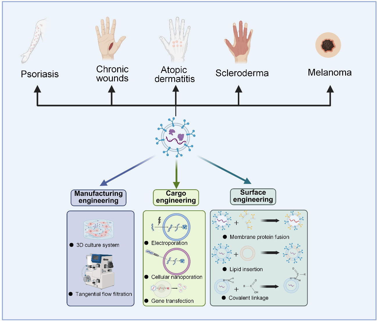
(Created with BioRender.com)
Skin diseases place a heavy burden on health-care systems worldwide. Chronic skin disorders are rarely life-threatening, but their long-term management continues to pose major clinical challenges [1]. These difficulties are partly related to the unique biological nature of the skin, which functions as an active immune interface continuously exposed to the external environment. In dermatologic disease, barrier dysfunction, immune activation, inflammation, and tissue repair are often interconnected and may change as the disease progresses [2, 3]. Established therapies involve the use of topical corticosteroids, systemic immunosuppressants, and targeted biologic drugs. These treatments can effectively ameliorate symptoms, but their prolonged use is limited by the risk of relapse, adverse effects, or incomplete restoration of skin homeostasis [4, 5]. As a consequence, investigators in this field are now searching for strategies that can facilitate the coordinated regulation of these interconnected processes.
Exosomes are nanoscale vesicles generated through the endosomal-multivesicular body pathway. They serve as natural carriers for bioactive cargo and mediators of intercellular communication [6]. Typically, 30–150 nm in diameter, these vesicles are enclosed by a phospholipid bilayer and carry nucleic acids, proteins and lipids derived from their parental cells [7]. Compared with many synthetic nanocarriers, native exosomes generally exhibit stability in physiological environments, favorable biocompatibility, and low immunogenicity [8-10]. Moreover, they offer practical advantages over cell-based therapies, including easier storage, repeated administration, and a lower theoretical risk of tumorigenicity [11]. A growing body of evidence supports their value in dermatology. Exosomes derived from skin cells, blood, and mesenchymal stem cells (MSCs) can promote angiogenesis and re-epithelialization while attenuating local inflammation, thereby supporting tissue repair in several experimental models [12-15]. Research interest has recently extended to plant-derived exosome-like nanoparticles (PDELNs), especially those isolated from traditional Chinese medicinal plants [16]. These PDELNs retain bioactive molecules from their source tissues and have shown antioxidant, anti-inflammatory, and tissue-repair activities [17]. Accessible sources and potential scalability render PDELNs even more attractive candidates for translational development.
Despite the therapeutic promise described above, native exosomes are not inherently suitable for efficient transdermal delivery. Their cargo composition is largely determined by the physiological state of donor cells, and consequently, targeting behavior, loading capacity, and batch consistency are often difficult to control [6, 18]. Engineering strategies have thus emerged to convert native exosomes into more programmable platforms. The methodological basis of this field can be traced to early work on cell-mediated drug packaging and targeted nucleic acid delivery. Pascucci et al. attempted to incorporate paclitaxel into secreted vesicles during exosome biogenesis, suggesting that MSCs could function as factories for effectively packaging antineoplastic molecules into vesicles [19]. A particularly interesting study was based on the fusion of the membrane protein lysosome-associated membrane protein 2b (LAMP2b) with targeting peptides. The results showed that this fusion protein could be transferred from donor cells to the exosomes they secreted and ultimately displayed on the vesicle surface [20]. Since then, interest in engineered exosomes has grown quickly and has gradually converged on two major directions: cargo loading to enhance bioactivity and surface modification to improve delivery specificity [21, 22]. For dermatologic diseases, such engineering is particularly important. Multifunctional vesicles are needed both to address interconnected pathological networks and to precisely target cells within inflammatory microenvironments. Further breakthroughs are required given that exosome populations are by nature heterogeneous, and this heterogeneity can compromise the production of stable therapeutic products. Therefore, scalable manufacturing, quality control, and process standardization are considered key priorities for clinical translation [7, 23].
Through engineering, exosomes can be transformed from natural intercellular messengers into more controllable and effective delivery platforms. This article seeks to systematically summarize the main modification techniques available, evaluate how these methods improve therapeutic performance, and highlight their applicability in treating representative dermatological diseases. By discussing current advances and remaining challenges, this review aims to provide a framework for understanding the role of engineered exosomes in the future development of precision dermatology.
To enable the therapeutic application of exosomes, it is necessary to determine which cargo they should carry, how they can be directed to their targets, and how they can be manufactured on a large scale.
2.1 Cargo engineering
The functionalization of exosomes is primarily achieved through cargo engineering. As depicted in Figure 1, cargo-engineering strategies can be categorized into endogenous loading, exogenous loading, and pre-formation loading. Pre-formation loading integrates cargo during exosome biogenesis and serves as a complementary engineering strategy.
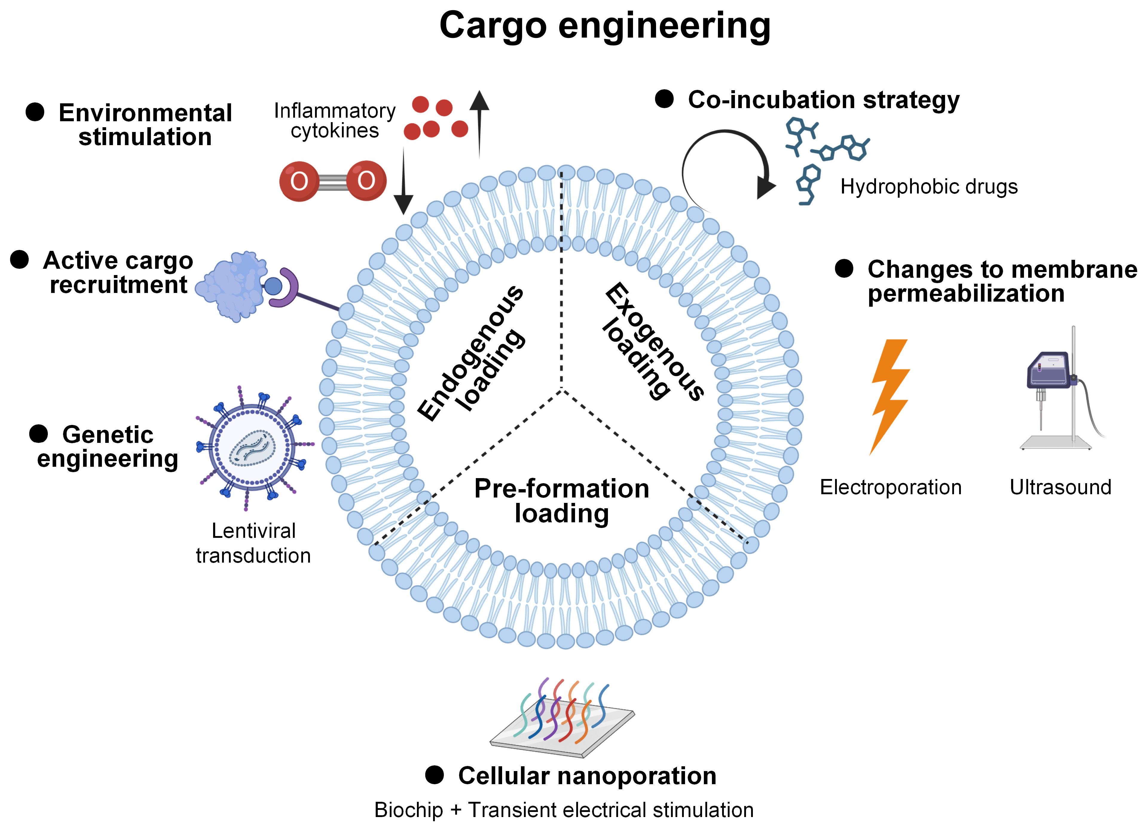
Figure 1. Cargo-engineering strategies for exosomes. Created with BioRender.com.
2.1.1 Endogenous loading
Exosomes loaded with a specific cargo are often generated through plasmid-based transfection or lentivirus-based transduction. This is possible because endogenous loading relies on exosome biogenesis rather than external physical methods. Cells overproduce the target cargo and package these molecules into exosomes [24]. Recent combinatorial designs have enabled the concurrent loading of nucleic acids and proteins. Yueh et al. clearly demonstrated this capability [25]. Moreover, fusing cargo with ubiquitin tags greatly increases protein loading [26]. Nevertheless, the abundance of molecules in the cytoplasm does not guarantee their incorporation into exosomes. The sorting ability of transmembrane proteins can help overcome this limitation [27, 28]. Passive diffusion alone is rarely sufficient for large cargo molecules. Large CRISPR-associated protein 9–single-guide RNA ribonucleoprotein complexes enter exosomes through a cluster of differentiation 63 (CD63)–MS2 coat protein (MCP) scaffold [29]. A strong interaction between the MCP domain and MS2 aptamers in the single-guide RNA drives the recruitment of editing tools. Later, tuning this MCP-MS2 interaction was shown to balance loading stability and intracellular release [30]. In this way, the system is able to incorporate different CRISPR-associated protein 9 variants without compromising efficiency.
In a low-oxygen environment, cells increase overall vesicle production, and the transcriptomic profile of their exosomes differs from that observed under normoxic conditions [31]. Energy metabolism shifts toward glycolysis, and angiogenesis is promoted. Such reactions are aimed at alleviating hypoxic stress across tissues [32]. Supplementation of the culture medium with melatonin enhances the regenerative properties of the resulting exosomes, as evidenced by elevated levels of the microRNAs let-7b-5p, miR-23a-3p, and miR-100-5p [33]. Exposure to tumor necrosis factor-α upregulates the autophagy-related protein 16-like 1, accelerating multivesicular body formation and, consequently, the packaging of the functional cargo [34]. These natural adaptive stress responses can be harnessed to produce engineered exosomes with an optimized payload landscape. In some cases, pharmacological stimulation can even induce exosomes to encapsulate entire mitochondria [35].
2.1.2 Exogenous loading
Curcumin, paclitaxel, and rapamycin can be easily loaded into isolated exosomes [8, 36-38]. Diffusion and hydrophobic interactions are the primary driving forces. This post-loading strategy simply involves incubating exosomes with these lipophilic molecules, which naturally partition into the lipid bilayer. However, it often suffers from low loading efficiency. Some vesicles are heavily loaded and others stay empty. Freeze-thaw cycles offer a low-cost approach, but due to the difficulty in maintaining membrane integrity, this technique has gradually fallen out of favor [39]. Active loading methods apply physical stimuli to transiently disrupt the membrane. Additionally, these direct-loading methods enable efficient processing. For example, electroporation or sonication can rapidly deliver larger amounts of cargo into exosomes, with loading efficiencies of up to 90% [40, 41]. Rather than relying on sorting by parent cells, such methods are relatively controllable. It is also possible to determine how much of the drug has successfully been encapsulated in the carrier. Following extrusion through a microporous membrane, the particles are nearly uniform in size [42]. The exosome membrane is transiently disrupted and then reseals, entrapping the drug inside.
There is emerging evidence that hybrid vesicles formed by combining exosomes with liposomes can deliver messenger RNA (mRNA) and peptide drugs [43, 44]. Fusogenic lipid nanoparticles termed cubosomes even load large therapeutic macromolecules into exosomes within minutes [45]. Pure exosomes generally lag behind synthetic nanocarriers in payload capacity. To overcome this limitation, hybrid vesicles merge the biocompatibility of exosomes with the physicochemical advantages of liposomes. Smaller and more uniform nanoparticles exhibit higher contact probability and undergo accelerated membrane fusion [46].
2.1.3 Pre-formation loading
To overcome low loading efficiency while avoiding damage to cell membranes, researchers have developed a series of strategies, one of which is cellular nanoporation (CNP) [47]. In this technique, cells cultured on a biochip are subjected to brief electrical pulses. Compared with bulk electroporation, CNP enables exosomes to accommodate large nucleic acids, leading to a more than 1,000-fold increase in exosomal mRNA transcripts. You et al. subsequently utilized CNP to deliver COL1A1 mRNA for photoaged skin and vascular endothelial growth factor A mRNA for ischemic injury, demonstrating a safer and more effective therapeutic strategy in both settings [48, 49]. In fact, this method still represents an improved form of genetic modification. Using a quantitative assay, RNA transfection efficiencies of 90–100% have been reported [50]. The term “pre-formation loading” was formally defined by Ramon et al., who employed photoporation to deliver cargo directly into the cytoplasm of producer cells [51]. As a result, different types of molecules can be encapsulated within the exosomes as they are assembled. Moreover, this flexible approach has minimal impact on vesicle characteristics.
2.2 Surface engineering
Surface engineering constitutes an equally critical strategy for optimizing exosome functionality and encompasses four primary approaches: membrane protein fusion, membrane insertion, chemical linkage, and metabolic labeling (Figure 2). These techniques equip exosomes to display targeting ligands or stealth molecules on their surface and fine-tune their interactions with recipient cells.
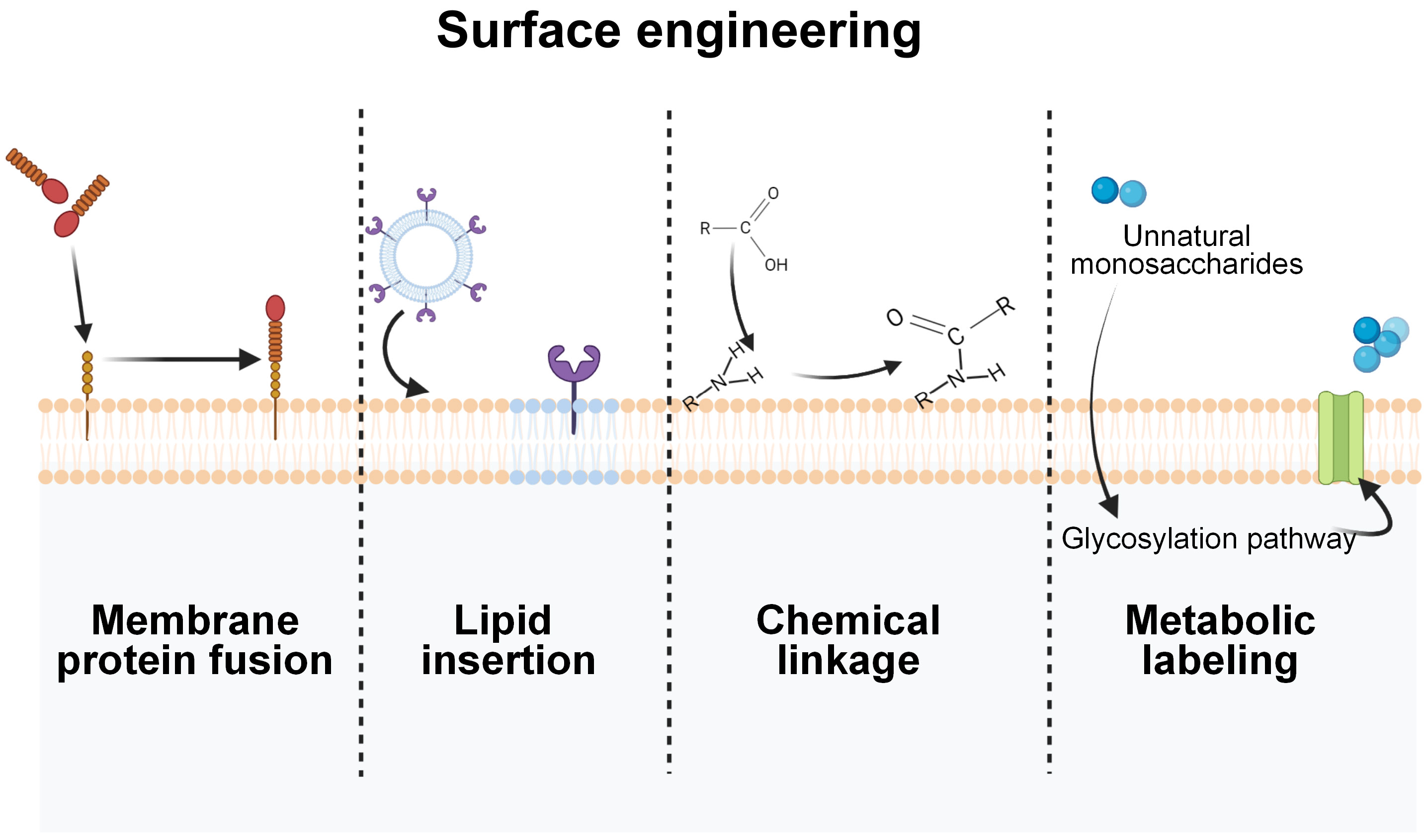
Figure 2. Surface-engineering strategies for exosomes. Created with BioRender.com.
2.2.1 Membrane protein fusion
Membrane protein fusion is a widely used genetic strategy for exosome surface engineering. By fusing functional peptides, ligands, or protein domains with exosomal membrane scaffolds such as CD63, CD9, CD81, and LAMP2b, donor cells can produce engineered exosomes displaying selected surface motifs that are readily accessible to receptors on recipient cells. This approach allows vesicles to acquire additional functions, including selective cell recognition, improved tissue penetration, immune regulation, and reduced clearance.
A CD63 fusion with a thrombopoietin receptor-binding peptide is able to selectively guide exosomes toward leukemia cells [52]. Similar principles have been applied to overcome biological barriers. Angiopep-2, a ligand associated with blood-brain barrier transport, can improve vesicle access to glioblastoma lesions, whereas TAT peptides enhance membrane penetration and cellular uptake [53, 54]. Programmed death-ligand 1 (PD-L1)-enriched exosomes confine immunosuppressive signaling to inflamed sites [55]. Owing to its favorable membrane topology, LAMP2b is preferred as a scaffold for the co-display of PD-L1 and galectin-9. This dual-ligand design suppresses excessive immune activation and further promotes the silencing or removal of activated effector T cells [56]. Immune evasion is also an important goal of membrane protein fusion. CD47 expressed on host cells binds to signal regulatory protein α on macrophages and limits phagocytic clearance. Exosomes can inherit this “don’t eat me” signal from parental cells, while those with enhanced CD47 presentation are advantageous for systemic delivery, repeated dosing, or the treatment of deep inflammatory lesions that require prolonged vesicle retention [57, 58].
However, the performance of membrane protein fusion depends strongly on the choice of scaffold. Conventional exosomal membrane scaffolds are constrained by their topology, orientation, and sorting efficiency. To address these limitations, Plexin A1 has been proposed as an alternative display scaffold [59]. Plexin A1 is a type I transmembrane protein and remains functional after truncation. Its N-terminal region provides flexible sites for ligand insertion, supporting complex surface designs.
2.2.2 Lipid insertion
Membrane insertion is a post-isolation engineering technique in which lipid anchors are inserted into the exosomal phospholipid bilayer. This approach is straightforward to implement and can modify vesicles under mild conditions, thereby preserving membrane integrity and fluidity. Hydrophilic ligands, aptamers, or small interfering RNA (siRNA) molecules are usually conjugated to lipid anchors for display on the exosome surface. For this purpose, 1,2-distearoyl-sn-glycero-3-phosphoethanolamine–polyethylene glycol (PEG) is an amphiphilic linker. The 1,2-distearoyl-sn-glycero-3-phosphoethanolamine–PEG–aptamer module has been developed by Zhao et al. to direct exosomes to CD31+EMCN+ vessels in diabetic wounds [60]. The PEG segment positions the aptamer at an appropriate distance from the vesicle surface, thereby facilitating CD31 recognition. Conversely, compact RNA nanostructures that integrate both targeting ligands and siRNA require shorter spacers to maintain structural integrity [61].
For membrane insertion, the choice of lipid moiety is critical, as it influences insertion efficiency and retention time. Among the available lipids, 1,2-dioleoyl-sn-glycero-3-phosphoethanolamine represents a well-balanced and adaptable option. This lipid anchor can be coupled with fluorescent probes, targeting peptides, or albumin-binding groups [62].
2.2.3 Chemical linkage
Chemical linkage takes advantage of reactive groups that are naturally exposed on the exosome surface. Targeting ligands can be covalently attached to the vesicle membrane via amino, carboxyl, or thiol groups on membrane proteins and lipids. This chemistry, however, is sensitive to reaction conditions. Without tight control over reaction time, pH, and activation states, there is a substantial risk of disrupting the native folding of membrane proteins. It is also important to minimize residual coupling reagents or by-products. Chemical conjugation usually provides more durable modification than lipid anchoring because the ligand is irreversibly attached and is less likely to dissociate.
Commonly employed reactions include carbodiimide-mediated amide bond formation and thiol-maleimide coupling. Carbodiimide coupling using 1-ethyl-3-(3-dimethylaminopropyl)carbodiimide and N-hydroxysuccinimide (NHS) is well established and frequently adopted for surface engineering. The carbodiimide reagent (─N=C=N─) activates free carboxyl groups on exosomal membrane proteins. It induces the formation of O-acylisourea intermediates, which then react with NHS to form a more stable NHS ester. The modified AS1411 ultimately displaces NHS through the nucleophilic attack of its primary amines on the NHS ester [63]. However, this method lacks site selectivity since carboxyl groups are widely distributed on the surface. As a result, the final product may contain a heterogeneous population of exosomes with variable ligand density and orientation. Thiol-maleimide coupling offers higher site selectivity. Free sulfhydryl groups are less abundant. This narrows the range of reactive sites but also limits the total modification capacity. To create more available thiols, disulfide bonds can be reduced with reagents such as tris(2-carboxyethyl)phosphine [64]. Since reductive treatment may disturb protein conformation, milder enzymatic methods have also been explored. Chen et al. reported that, at physiological pH, phospholipase D exposed hidden thiol sites, enabling conjugation with maleimide-functionalized alcohols and achieving modification efficiencies above 90% [65].
2.2.4 Metabolic labeling
Surface modification is valuable only when it does not erase the biological identity of exosomes. Although the post-isolation methods mentioned above are convenient, they may still interfere with membrane structure or surface proteins to some extent. Metabolic glycoengineering, also referred to as metabolic labeling, addresses this limitation by shifting the modification process upstream to the donor cells. Instead of genetic decoration, cells are supplied with unnatural monosaccharide precursors, which enter the glycosylation pathway and are incorporated into newly synthesized glycans. The secreted exosomes therefore inherit bioorthogonal chemical handles on their surface. Azide groups (–N3) and alkynes, particularly strained alkynes such as dibenzocyclooctyne, are commonly selected for this purpose [66]. They are largely absent from native biomolecules and remain relatively inert under physiological conditions. Chen et al. applied this principle by using azide-labeled exosomes as a clickable scaffold for the conjugation of dibenzocyclooctyne-modified dextran sulfate [67]. This modification improved targeting to pro-inflammatory M1 macrophages and promoted macrophage polarization toward an M2-like phenotype.
Notably, metabolic labeling should not be regarded as completely passive. As Agatemor et al. discussed in their review, the competition between unnatural sugar substrates and endogenous sugars may alter cellular metabolism. They also suggested that unlabeled controls and natural sugar rescue experiments are useful for distinguishing labeling effects from broader metabolic perturbations [66].
2.3 Manufacturing engineering
2.3.1 Cell sources and three-dimensional (3D) culture
Manufacturing engineering begins with the producer cell. All functionally viable cells secrete exosomes. Natural sources, such as plants or biological fluids, have clear advantages in production and collection. MSCs remain a major research focus in the treatment of skin diseases, as the regenerative and immunomodulatory effects of their exosomes are difficult to replicate. The challenge is that primary mammalian cells have limited expansion capacity, and their characteristics and functions change during prolonged culture. Therefore, immortalized cell lines have been explored. Tong et al. introduced simian virus 40 large T antigen into human placental chorionic MSCs, achieving long-term expansion for over 50 passages [68]. This antigen disrupts cell-cycle regulation and places cells in a less physiologically relevant proliferative state. Alternatively, human telomerase reverse transcriptase-based immortalization, which extends cellular lifespan through telomere maintenance, may raise fewer concerns regarding oncogenicity [69]. Exosomes from these immortalized cells, even after several passages, showed similar proteomic profiles to those from primary cells.
The culture method is equally important for improving exosome yield. Conventional two-dimensional culture is easy to implement, but it is not suitable for sustained production. Cells grow on a flat surface and their growth soon becomes constrained by the available area. In contrast, 3D culture overcomes this spatial limitation. Adherent cells grow on suspended microcarriers rather than on the bottom of culture dishes [70]. This greatly increases production within a short time. To produce 1012 particles, one microcarrier system required only a single shake flask and 488 mL of conditioned medium [71]. More advanced 3D systems using flexible substrates and microgravity culture are designed to better mimic in vivo tissues [72, 73]. These methods may help prevent the reduction in exosome release associated with excessive cell spreading. In microfluidic chips, fluid shear stress can be controlled to stimulate secretory activity [74, 75].
2.3.2 Isolation and purification
Downstream isolation is a decisive step in exosome manufacturing. The aim is not simply to collect more vesicles, but to remove soluble proteins, protein aggregates, lipoproteins, and other particles.
Differential ultracentrifugation has long been regarded as a classical laboratory method. Repeated centrifugation and resuspension can improve purity, but also make the workflow time-consuming and difficult to scale. Thus, centrifugation-based methods are useful for concentration and initial enrichment, but they are not ideal as stand-alone methods for manufacturing [76]. For larger sample volumes, filtration is more practical. Tangential flow filtration is particularly effective for plant-derived samples, where fibrous residues and polysaccharides may accelerate membrane fouling. Still, filtration mainly retains vesicles based on a molecular weight cutoff, so protein aggregates, lipoproteins, and other particles of similar dimensions may also be retained in the final product. It is therefore commonly combined with size exclusion chromatography, a gentle method that further separates exosomes from soluble contaminants [77]. Affinity capture enriches exosomes through defined surface markers, making it suitable for well-characterized exosome subpopulations or genetically engineered exosomes carrying specific tags [78].
Recently, several emerging platforms have been developed to simplify operation. Photosensitive lipid nanoprobes, for example, exploit the biotin-avidin interaction to enable labeling, capture, and release within a short workflow [79]. Ion chromatography employs the negative charge of phospholipids to bind exosomes to the stationary phase, and has been applied to the rapid processing of liquid biopsy samples [80, 81]. Exosome detection via the ultrafast-isolation system represents another automated platform capable of isolating exosomes from diverse biological fluids [82]. Even so, the value of these technologies for exosome manufacturing should not be judged by novelty alone. Scalability, cost, sample compatibility, release efficiency, and preservation of vesicle function all need to be considered.
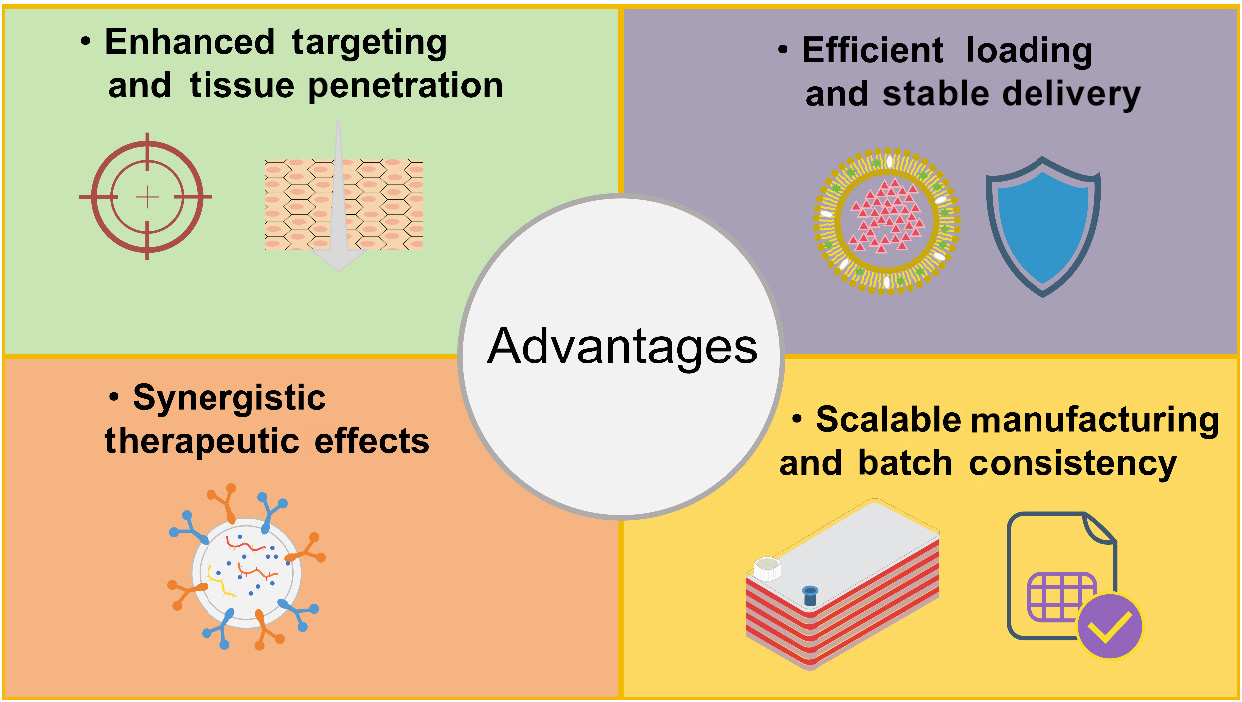
Figure 3. Advantages of engineered exosomes in dermatology. Created using Microsoft PowerPoint.
3.1 Enhanced targeting and tissue penetration
In some skin diseases, pathological changes are located deep within the dermis. Some stem cells and their exosomes possess homing properties. However, this natural tendency is insufficient when specific interactions with certain target cells are required [11]. Surface engineering addresses this limitation by exploiting receptor-ligand interactions. Vesicles enriched with PD-L1 can directly inhibit T cells [55]. Synthetic aptamers were used by Zhao et al. to label exosomes, increasing the binding selectivity of the exosomes toward endothelial cells by more than tenfold [60]. Similar ideas appear in other surface modification strategies. Metabolic glycoengineering or membrane fusion, for instance, endows engineered exosomes with higher affinity for particular macrophage subpopulations [67, 83]. Some engineering designs incorporate stimulus-responsive elements rather than relying on cell-type recognition. pH-sensitive peptides stay quiescent under physiological conditions but become activated in the acidic microenvironment. These peptides are anchored on the surface of exosomes, so that cargo release mainly occurs at tumor sites or sites of active inflammation [84, 85].
Nevertheless, if the engineered exosomes fail to enter the skin, the value of targeting becomes limited. The stratum corneum functions as a strong barrier. Exosomes may cross it through hair follicles or along intercellular lipid channels. Some evidence supports this possibility, but when the skin is intact, the contribution of these routes to penetration is relatively small [86, 87]. Among invasive techniques, microneedle platforms are frequently used to deliver vesicles deep into tissues [88]. Li et al. aimed to enable light-controlled and deep transdermal delivery, so they developed a spherical nucleic acid that integrates gas-generating molecules [89]. The 3D nanostructure can penetrate to a depth of up to 120 μm into the dermis.
3.2 Efficient loading and stable delivery
Free drugs are frequently degraded in the body. Exosomes have a relatively stable lipid bilayer structure, which protects the cargo inside. For instance, anthocyanins are highly sensitive to environmental conditions. When such unstable compounds are encapsulated within vesicles, their retention is much higher than that of the corresponding free compounds [39]. Drug-loaded vesicles represent a major category of engineered exosomes. Besides maintaining product quality, cargo engineering also seeks to enhance efficacy. The employment of an engineered CD63 scaffold makes autonomous cargo selection possible [90]. Compared to simple passive loading, the ultrasonic incubation method has been reported to raise loading efficiency to around 85% [91].
Loading the cargo is only one part of the process. The more critical question is whether the payloads are released once exosomes enter the target cells. Several factors influence this step, including temperature and pH, both of which can affect cellular uptake behavior [92]. Even after exosomes are internalized, delivery is not yet complete. The cargo still has to evade lysosomal degradation before it can eventually reach the cytoplasm [93]. Avoiding lysosomal degradation is one of the main obstacles to the application of natural exosomes. Some engineering strategies have been proposed for this stage. Vesicular stomatitis virus glycoprotein promotes membrane fusion between exosomes and endosomes. Through the resulting fusion pore, mRNA contained in the vesicle lumen escapes into the recipient cell [27]. At the same dose, these engineered exosomes enable nearly complete cytoplasmic delivery of mRNA. The lipid composition of the exosomal membrane also influences this process [94]. Certain lipid metabolic signatures have been found to promote macrophage uptake of anti-inflammatory exosomes [95]. Further analyses suggest that palmitoylation of membrane lipids is closely associated with the membrane anchoring of fusion proteins [96]. Once inside the endosome, such palmitoylated lipids may increase the probability of triggering membrane fusion.
3.3 Synergistic therapeutic effects
One approach is to modify the genetic program of donor cells to alter their secretory profile. The exosomes produced in this way acquire additional functional molecules. Vesicles rich in the transcriptional repressor Foxp1 promote the conversion of conventional T cells into regulatory T cells (Tregs) [102]. Stem cells are engineered to overexpress interferon regulatory factors. This change in turn leads to elevated levels of miR-16-5p in the released exosomes. Target cells take up miR-16-5p, which gradually alleviates the inhibitory effect on wound healing [103]. Another type of exosome carries cytotoxic molecules that induce apoptosis in melanoma cells. After loading these vesicles with chlorin e6, researchers utilized their photoresponsive behavior for precise photodynamic elimination of tumor cells [104].
In the treatment of skin diseases, exosomes are generally used for local immunomodulation, making it particularly difficult for them to rapidly suppress acute, severe inflammatory storms. Conventional dermatological drugs are potent but non‑specific. Dexamethasone is a representative example, but relapse after drug withdrawal and adverse effects remain concerns. The combination of dexamethasone and exosomes was demonstrated by Ma et al. to exert a synergistic biological effect against systemic lupus erythematosus [105]. Dimethyl fumarate is suitable for adult patients with moderate to severe psoriasis. Treg exosomes loaded with dimethyl fumarate not only alleviate skin symptoms, but also overcome immune dysfunction. This comprehensive approach is superior to either component deployed individually [106].
3.4 Scalable manufacturing and batch consistency
Clinical translation of engineered exosomes requires more than simply scaling up production volume. The final product must also remain consistent in terms of identity, potency, and safety. Many early-stage studies still depend on two-dimensional culture and differential ultracentrifugation, which are suitable for exploratory experiments but difficult to adapt to mass production. In static mammalian cell culture, the restricted growth area requires frequent monitoring and adjustment of cell state. As a result, the secretory profile of producer cells may vary between batches with changes in cell density, nutrient consumption, and harvest timing [107]. For plant-derived exosomes, additional variables arise during sample pretreatment. When plant homogenates are processed by ultracentrifugation, mechanical stress may lead to variable vesicle loss. Fluctuations that seem acceptable at laboratory scale can become amplified during scale-up.
Industrial experience with biological products offers a more practical direction for exosome manufacturing. Perfusion culture maintains a continuous flow of fresh medium and enables vesicles to be harvested soon after secretion [108]. This shortens their residence time in the bioreactor and may minimize aggregation or degradation. When tangential flow filtration is integrated with bioreactors, a closed and automated production workflow can be established [109, 110]. Once key parameters are defined in advance, each batch is more likely to follow a consistent and stable production pattern, with minimal variation introduced by external factors. In this sense, clinical scalability depends not only on production capacity, but also on whether the process can repeatedly generate vesicles with comparable quality.
Diverse pathological mechanisms drive the development of chronic skin diseases, ranging from immune dysregulation and fibrotic abnormalities to impaired tissue repair. However, they share a common imbalance in the microenvironment, primarily due to the dysregulation of interconnected signaling networks. Engineered exosomes, as a customizable platform, can meet the needs of different pathological stages.
4.1 Psoriasis
The interleukin-23/T helper 17 cell signaling pathway plays a central role in psoriasis, and its influence extends well beyond immune activation, as it also triggers extensive immunometabolic dysregulation [111-113]. Rapid keratinocyte proliferation follows chronic inflammation, while the lipid metabolites generated in the process further fuel inflammation [114]. Elevated glycolysis supports the pathogenic differentiation of effector T cells and macrophages [115]. Current biologic therapies mainly focus on single cytokines such as interleukin-17, interleukin-23, or tumor necrosis factor-α. These therapies control only parts of the inflammatory cascade. Such treatments lack metabolic regulatory functions [116, 117].
The coexistence of immune dysregulation and metabolic reprogramming has motivated the use of engineered exosomes as multifunctional delivery platforms. One strategy focuses on polyamine metabolism [118]. MSC-derived exosomes have been engineered to carry an arginase-1 inhibitor that depletes precursors for polyamine synthesis (Figure 4A). Autoantigen levels and the activity of related antigen-presentation pathways are markedly reduced. Researchers have also attempted combinatorial loading to influence several pathways at the same time. Ultraviolet B exposure upregulates platelet-activating factor in keratinocytes, thereby transmitting immunosuppressive signals to neighboring keratinocytes [119]. This effect can be enhanced by loading exosomes with JPH203, an L-type amino acid transporter 1 inhibitor [120]. Excessive activation of the mechanistic target of rapamycin (mTOR) is a major factor contributing to keratinocyte proliferation. Extracellular leucine turns on this cascade by binding to sestrin2 and inducing its dephosphorylation [121]. JPH203 can occupy the binding site, thereby hindering leucine transport. Exosomes also serve as carriers for natural bioactive molecules from medicinal plants, including epigallocatechin-3-gallate and anhydroicaritin [122, 123]. The lipid bilayer protects these molecules from rapid degradation, reducing the skin irritation that may occur during topical application.
Another advantage is that engineered exosomes can precisely target inflamed lesions. Damaged tissue releases chemotactic signals that recruit reparative exosomes, reflecting the body’s natural compensatory response. In particular, vesicles derived from MSCs have intrinsic homing properties, which facilitate the delivery of antisense oligonucleotides targeting miR-210 to CD4+ T cells, thereby regulating the balance among CD4+ T-cell subtypes [11, 124]. Liu et al. chose melanoma cells as the source because these cells naturally express PD-L1, the ligand for programmed cell death protein 1 (PD-1) on T cells [125]. Genetic engineering can further increase the amount of PD-L1 carried by exosomes, so that significant therapeutic effects can be achieved at lower doses (Figure 4B) [55].
Psoriasis lesions are accompanied by epidermal hyperplasia and hyperkeratosis, and the thickened plaques that form make drug penetration challenging [112]. Microneedle-assisted systems can penetrate this barrier, allowing engineered exosomes to enter the skin through the resulting micropores. Dissolvable microneedle patches are more convenient and less invasive [106]. Traditional phototherapy for psoriasis has poor efficacy, largely because the epidermis intercepts and absorbs much of the light. An optical microneedle system constructed by Zhao et al. guided the energy directly to the deep subcutaneous tissues [126]. Under controlled light exposure, it additionally released anti-inflammatory exosomes in the same vicinity (Figure 4C).
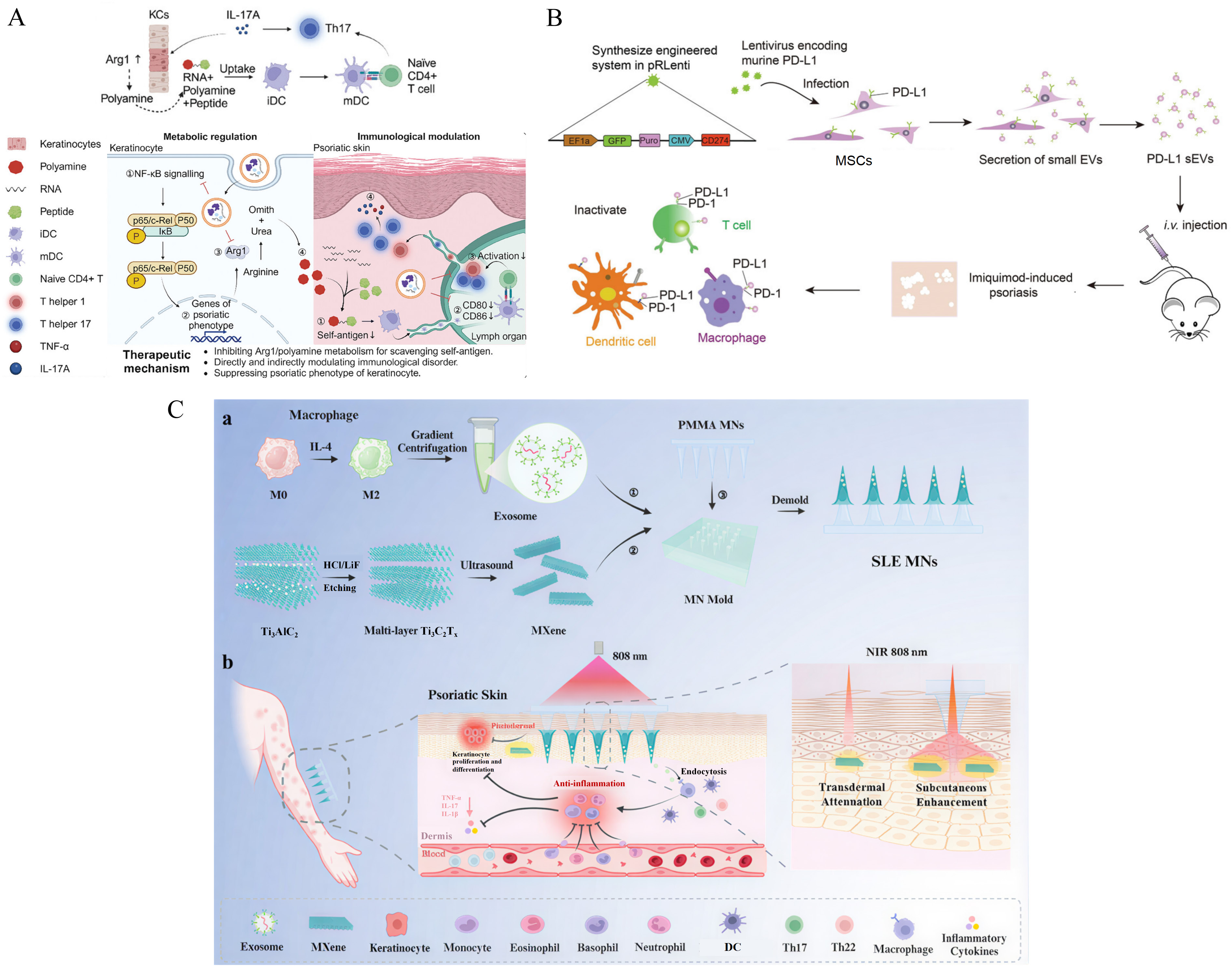
Figure 4. Engineered extracellular vesicle-based strategies for psoriasis therapy. (A) A synergistic strategy for regulating polyamine metabolism and the immune microenvironment, in which engineered MSC-derived extracellular vesicles disrupt the metabolic–immune vicious cycle in psoriasis. Adapted from [118] (Copyright © 2024, Zhou et al. Advanced Science published by Wiley‐VCH GmbH); (B) MSC-derived small extracellular vesicles with high PD-L1 expression inhibit the activation of T cells, macrophages, and DCs. Adapted from [55] (Copyright © 2021, Xu et al. Advanced Materials published by Wiley‐VCH GmbH); (C) A light-responsive microneedle system integrating immunomodulation with transdermal near-infrared light delivery. Adapted from [126] (Copyright © 2025, Zhao et al. ACS applied materials & interfaces published by ACS). Arg1, arginase 1; CD4, cluster of differentiation 4; CD80, cluster of differentiation 80; CD86, cluster of differentiation 86; CD274, cluster of differentiation 274; CMV, cytomegalovirus; DC, dendritic cell; EF1α, elongation factor 1 alpha; EV, extracellular vesicle; GFP, green fluorescent protein; HCl, hydrochloric acid; iDC, immature dendritic cell; IL-4, interleukin-4; IL-17A, interleukin-17A; i.v., intravenous; IκB, inhibitor of nuclear factor κB; KC, keratinocyte; LiF, lithium fluoride; M0, unpolarized macrophage; M2, alternatively activated macrophage; mDC, mature dendritic cell; MN, microneedle; MSC, mesenchymal stem cell; NF-κB, nuclear factor κB; NIR, near-infrared; PD-1, programmed cell death protein 1; PD-L1, programmed death-ligand 1; PMMA, poly(methyl methacrylate); RNA, ribonucleic acid; sEV, small extracellular vesicle; SLE MN, subcutaneous light response-enhanced microneedle; Th2, T helper 2 cell; Th17, T helper 17 cell; p65, RELA p65 subunit; p50, nuclear factor κB1 p50 subunit; c-Rel, REL proto-oncogene, NF-κB subunit; Puro, puromycin-resistance marker; Ti3AlC2, titanium aluminum carbide; Ti3C2Tx, titanium carbide MXene with surface terminations; MXene, a class of two-dimensional transition-metal carbides and nitrides; pRLenti, lentiviral expression vector.
4.2 Atopic dermatitis (AD)
AD is characterized by recurrent episodes and an immune response dominated by T helper 2 (Th2) cells [127, 128]. Due to immune bias, even some harmless stimuli can activate Th2 cells, which then release large amounts of interleukin-4 and interleukin-13 that aberrantly inhibit the normal synthesis of ceramides, cholesterol, and fatty acids. The “brick-and-mortar” structure of the skin becomes disrupted, facilitating the penetration of exogenous allergens. Breaking this positive feedback loop requires targeting multiple pathogenic factors: topical moisturizers strengthen the skin barrier, while abrocitinib or dupilumab block inflammatory signals.
Hereditary filaggrin deficiency is a major genetic risk factor [129, 130]. This could partly explain the high prevalence of AD observed in early childhood. When the filaggrin gene was knocked out, keratinocytes released altered exosomes with an abnormal lipid composition [131]. Such vesicle changes are often present in AD patients, and interleukin-13 levels are likely to be elevated. These findings suggest exosomes may be part of a vicious circle. As a consequence, exogenously introducing therapeutic exosomes seems to be a reasonable strategy. Evidence suggests that this approach may provide both anti-inflammatory effects and tissue repair [132, 133]. Optimizing the cell source further improves their potential. MSCs can be licensed by inflammatory cytokines to function in a highly activated state. Interferon-γ is a well-documented and stable licensing factor [134]. In experiments by Kim et al., exosomes obtained from interferon-γ-primed MSCs suppressed the expression of interleukin receptors for Th2 cytokines, including interleukin-4 receptor α, interleukin-13 receptor α1, and interleukin-31 receptor α [135]. The damaged skin barrier was restored, as evidenced by the recovery of key genes involved in epidermal differentiation and lipid synthesis from inhibitory effects (Figure 5A, 5B). Moreover, engineered exosomes are being redefined as versatile delivery platforms. Given its associations with improved sleep quality, skin cell protection, and reduced serum immunoglobulin E levels, a transdermal exosome-based delivery system for melatonin could be beneficial for AD therapy [136]. Attention is increasingly turning to cross-species membrane fusion, a novel, low-cost approach to integrating the benefits of distinct species. Easily obtainable exosomes—from sources such as Portulaca oleracea L., turmeric, or grapefruit—are known to alleviate symptoms of AD [137-139]. When such vesicles are fused with stem cell membranes, the resulting hybrid vesicles preferentially accumulate in regions enriched with chemokines [139]. RNA polymerase I inhibitors are then released to increase the proportion of Tregs within the inflamed lesion (Figure 5C).
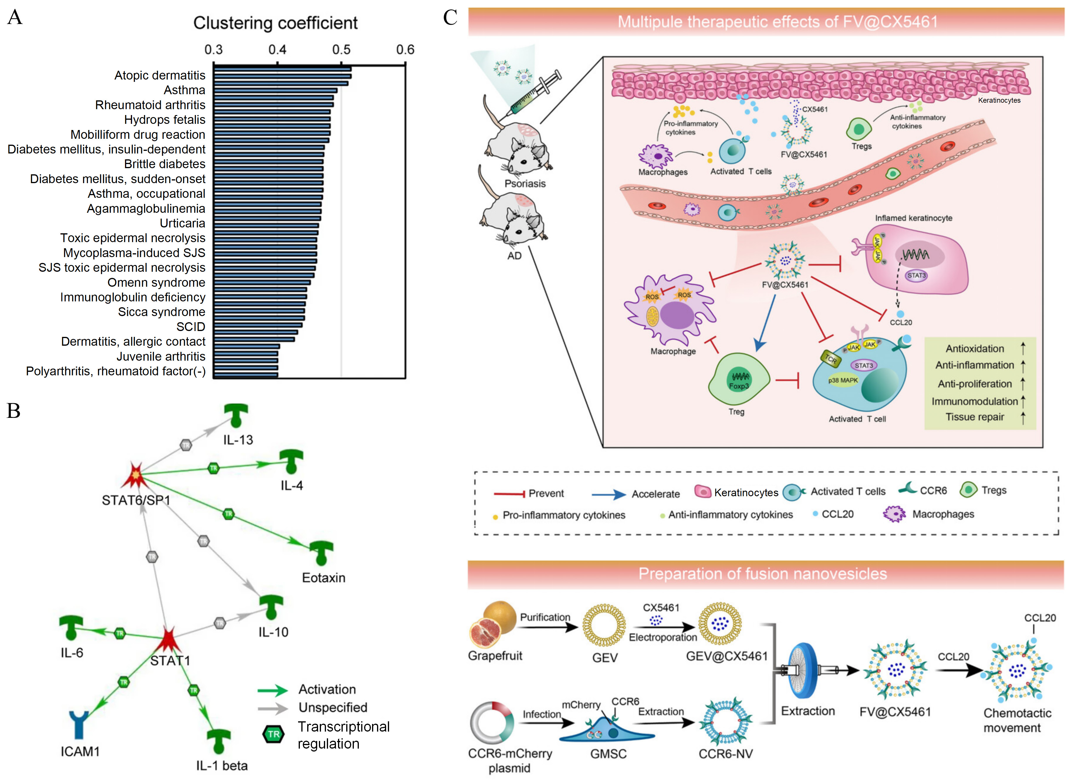
Figure 5. Engineered exosomes in atopic dermatitis. (A) Differential proteins identified in engineered exosomes show a significant association with the AD-associated gene set. Adapted from [135] (Copyright © 2022, Kim et al. Journal of Nanobiotechnology published by Spring Nature); (B) Engineered exosomes ameliorate Th2-type inflammation and downstream pathology in AD by modulating the IL-4/IL-13-STAT6/STAT1 signaling axis. Adapted from [135] (Copyright © 2022, Kim et al. Journal of Nanobiotechnology published by Spring Nature); (C) Design and function of the FV@CX5461 hybrid vesicle. Adapted from [139] (Copyright © 2023 Huang et al. Journal of Extracellular Vesicles published by Wiley Periodicals LLC on behalf of International Society for Extracellular Vesicles). AD, atopic dermatitis; CCL20, C-C motif chemokine ligand 20; CCR6, C-C motif chemokine receptor 6; FV, fusion vesicle; GEV, grapefruit-derived exosome-like nanovesicle; GMSC, gingiva-derived mesenchymal stem cell; ICAM1, intercellular adhesion molecule 1; IL-1β, interleukin-1β; IL-4, interleukin-4; IL-6, interleukin-6; IL-10, interleukin-10; IL-13, interleukin-13; MS, multiple sclerosis; NV, nanovesicle; SCID, severe combined immunodeficiency; SJS, Stevens–Johnson syndrome; SP1, specificity protein 1; STAT1, signal transducer and activator of transcription 1; STAT6, signal transducer and activator of transcription 6; Th2, T helper 2 cell; Treg, regulatory T cell.
4.3 Scleroderma
Systemic sclerosis (SSc), at its core, is a disease of progressive fibrosis [140]. Inflammation and vasculopathy are the initial events, but extensive damage results from uncontrolled fibroblast activation. These activated fibroblasts produce excessive extracellular matrix, disrupting homeostasis and leading to fibrosis and stiffening of the skin and internal organs [141, 142].
Small RNA profiling of circulating exosomes isolated from blood shows downregulation of anti-fibrotic microRNAs and upregulation of pro-fibrotic microRNAs [143]. Accordingly, unaffected cells may take up these pathological exosomes and acquire a fibrotic phenotype. Wermuth et al. instead focused on fingertip ulcers, a complication of SSc. They found that serum exosome levels were decreased in patients with SSc, possibly because of severe microvascular injury [144]. Only a small number of exosomes are released from dermal fibroblasts, whereas the secretion of inherently reparative exosomes is compromised. This hypothesis was supported in a mouse model in which normal serum-derived exosomes were applied to full-thickness skin wounds. Platelets may be the main contributors. In the context of SSc, natural exosomes present both challenges and therapeutic opportunities that warrant further investigation. Rozier et al. provided evidence that miR‑29a‑3p from MSC-derived exosomes can target genes related to apoptosis and methylation [145]. DNA methyltransferase 3 alpha, in particular, plays a critical role in epigenetic dysregulation during disease progression. Jin et al. identified miR‑143‑3p as a critical antifibrotic factor and demonstrated that miR-143-3p-enriched extracellular vesicles represent a potential therapeutic strategy for localized scleroderma [146]. Another important finding from their study is that this microRNA can be loaded into exosomes at high levels to more effectively inhibit extracellular matrix deposition, fibroblast differentiation, and epithelial cell activation.
4.4 Chronic wound healing in diabetes
Chronic wound healing continues to be a significant clinical issue. This is especially true for patients with metabolic disorders [147]. In diabetes, hyperglycemia disrupts cell function, predisposing patients to chronic ulcers, most commonly diabetic foot ulcers (DFUs). Acute inflammation is a beneficial early stage for normal wound repair. However, DFUs often remain in a state of low-grade but persistent inflammation. The accumulation of advanced glycation end products and reactive oxygen species traps macrophages in an inflammatory M1 state [148]. This inflammatory environment is exacerbated by protease imbalance. Abnormally elevated matrix metalloproteinases rapidly degrade growth factors and matrix [149]. If left uncontrolled, these alterations eventually lead to severe microvascular dysfunction and tissue hypoxia.
Wound healing typically goes through four phases. Unlike acute wounds, chronic ulcers bypass the hemostatic stage because platelets fail to aggregate and initiate the repair cascade. Platelet-rich plasma appears attractive, and it is frequently employed in regenerative medicine. When neutrophils take up platelet-rich plasma-derived exosomes, the suppressive miR-26b-5p is released to target matrix metalloproteinase-8, an important enzyme involved in the formation of neutrophil extracellular traps [150]. Damage-associated molecular patterns are attenuated, creating a favorable microenvironment for wound healing. Moreover, exosome therapy promotes the crosstalk between keratinocytes and macrophages. Sharma et al. found that macrophages enriched in the mitochondrial membrane protein TOMM70 often localize to leading-edge keratinocytes to rescue their mitochondrial metabolism [151]. In turn, keratinocytes deliver exosomal microRNAs to M1 macrophages, signaling that inflammation has already resolved [152].
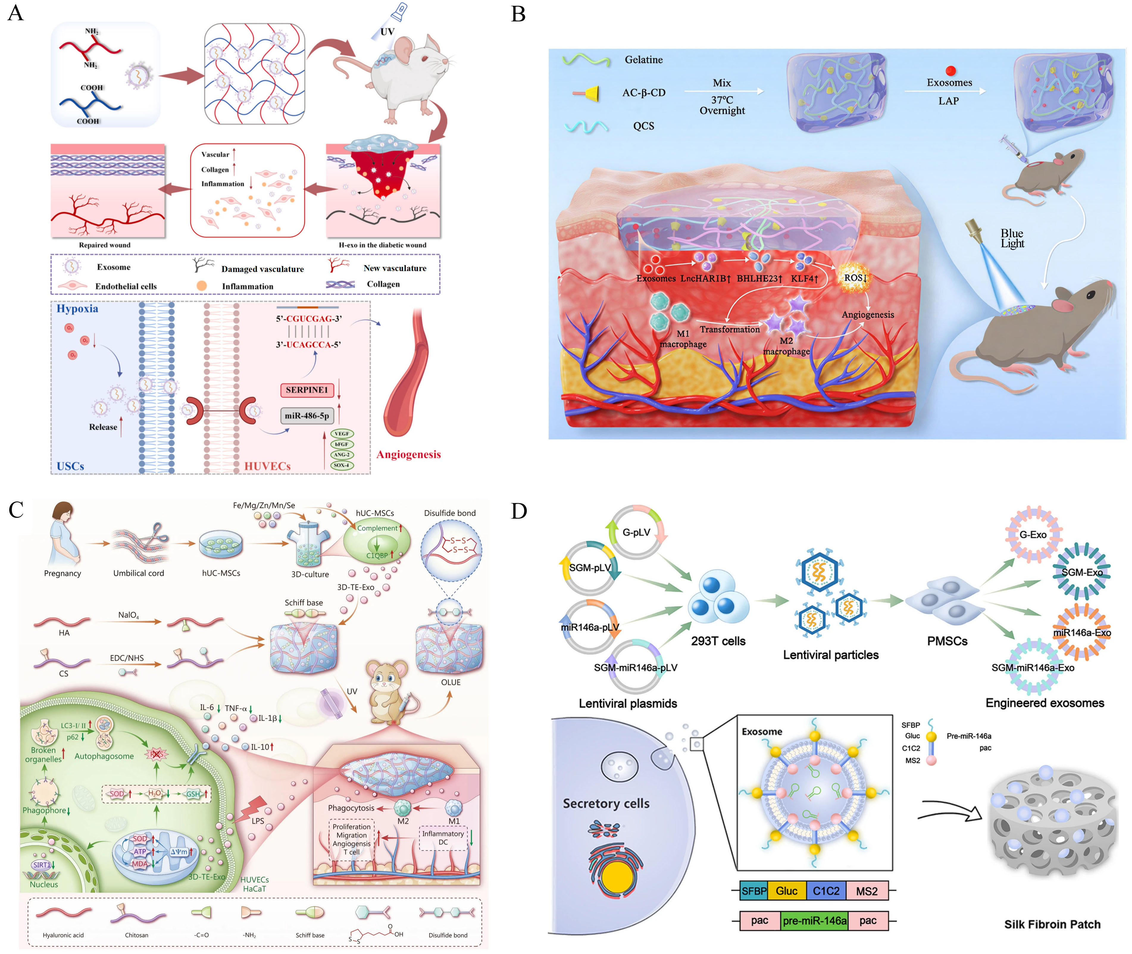
To better counteract pathological cascades in DFUs, engineering strategies have been applied to enhance exosomal efficacy. Stem cells have excellent regenerative capabilities. Those isolated from hypoxic tissues are more likely to maintain their stemness. Hypoxic preconditioning shifts the microRNA composition of exosomes, resulting in the upregulation of miR-21-3p, miR-126-5p, and miR-31-5p [153]. Fan et al. uncovered an association among exosomal miR-486-5p, the negative regulator of angiogenesis SERPINE1, and the hypoxia-inducible factor 1α signaling pathway (Figure 6A) [154]. In addition, differentially expressed long non-coding RNAs have been identified in exosomes from human umbilical vein endothelial cells. Cheng et al. ultimately pinpointed long non-coding RNA HAR1B, which exerts specific dual functions rather than broad-spectrum regulation (Figure 6B) [155]. Other types of nucleic acids, circular RNAs, work mainly as “molecular sponges”. Once inside diseased cells, they sequester microRNAs that would otherwise block repair-related gene expression. Thus, downstream genes, like those involved in hypoxia response and growth regulation, can be restored [156, 157]. Adding trace elements to a 3D dynamic culture system is also a method for manufacturing highly active exosomes (Figure 6C), which target multiple pathways through a complement–mitochondria–autophagy loop [158]. IMMUNEPOTENT CRP has recently been considered for DFU treatment, and its exosomes indeed promote tissue regeneration, with targets including but not limited to the phosphatidylinositol (3,4,5)-trisphosphate/protein kinase B signaling pathway [159]. IMMUNEPOTENT CRP-derived exosomes can also work alongside conventional drugs (insulin, gentamicin, etc.) to improve the wound microenvironment and help manage infection [160, 161].
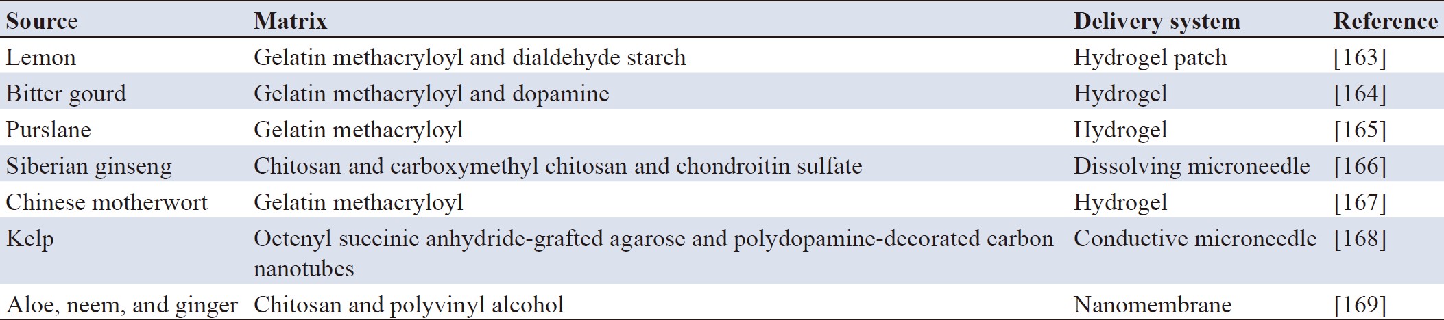
Since diabetic wounds are rich in proteases, clinical formulations are mostly prepared using hydrogels, microneedles, or other biomaterial platforms, such as silk fibroin [162]. The enzymatic degradation of this material is relatively slow, and when exosomes are anchored to such a silk fibroin patch via binding peptides, they can be released in a sustained and stable manner as the matrix degrades (Figure 6D). In recent years, exosomes intended for treating diabetic ulcers have been increasingly derived from medicinal or edible plants (Table 1).
4.5 Melanoma
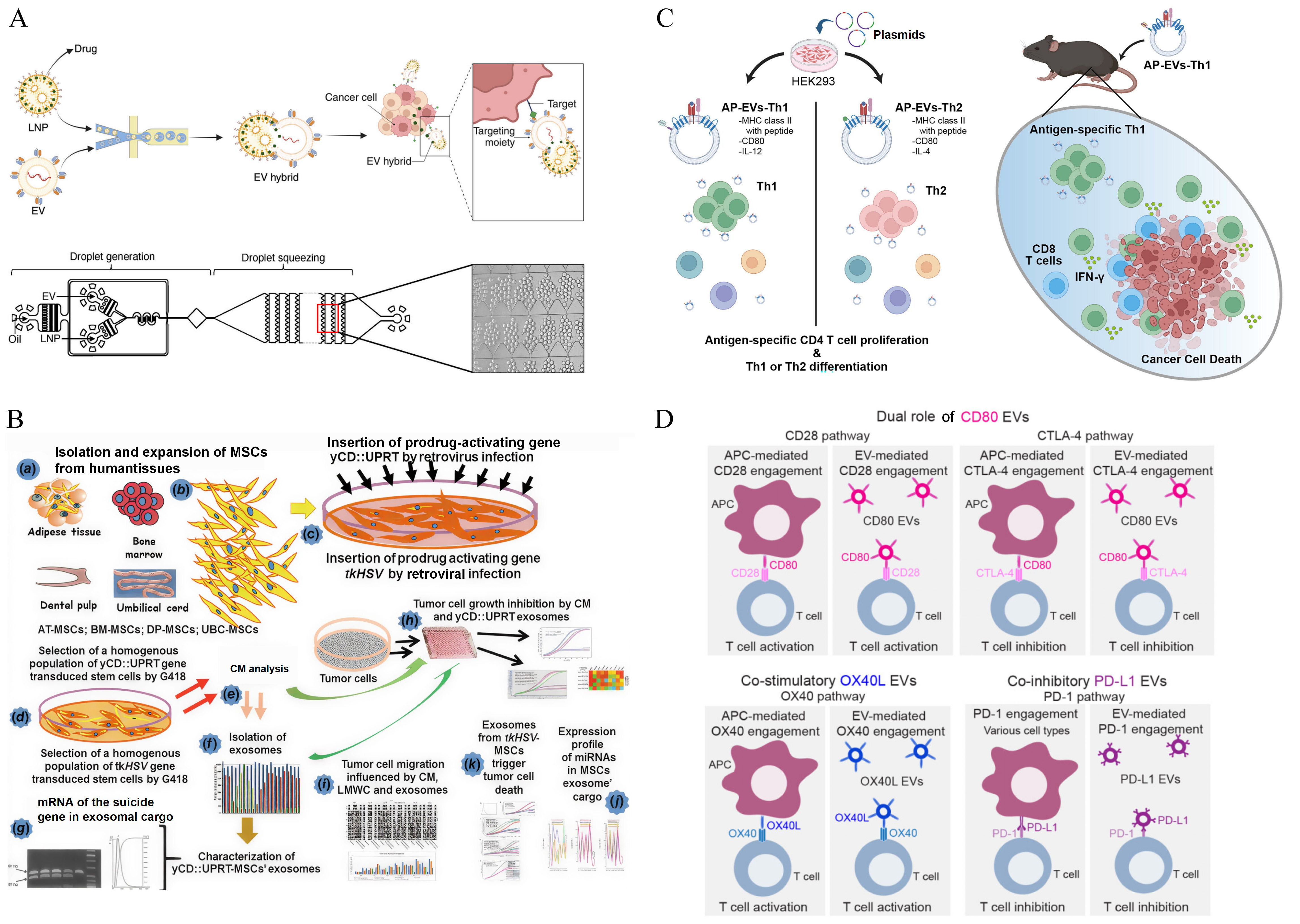
Skin cancer accounts for only a small proportion of cancer-related mortality, and most cases are relatively indolent types, such as basal cell carcinoma and squamous cell carcinoma, which originate from epidermal cells. Melanoma, however, is notably aggressive [170]. Its survival outcomes are even poorer than those of many advanced visceral cancers, largely due to its high metastatic potential. Some tumor cells can rapidly penetrate the basement membrane and seed the dermis at an early stage. This vertical invasion may be attributed to the epithelial-mesenchymal transition. A subset of macrophages (APOE+CD163+) was considered a potential initiator, and Liu et al. identified four invasion-associated driver genes: NRAS, KRAS, NF1, and KIT [171]. Mutant KRAS often activates the mitogen-activated protein kinase signaling pathway. Downstream transcription factors then bind to the death receptor 5 (DR5) promoter, resulting in high DR5 expression on the surface of tumor cells [172]. A single-chain variable fragment is a versatile antibody fragment that can be engineered as DR5-single-chain variable fragment to specifically target this antigen. A single immunoglobulin G molecule has only two binding sites, and its binding is stochastic. Exosomes aggregate on the membrane of DR5+ melanoma cells, effectively inducing conformational changes in the intracellular death domain of DR5 [173]. To meet the demand for clinical applications, Chintapula et al. scaled up production using microfluidic technology [75]. They additionally loaded extracellular signal-regulated kinase inhibitors to enhance the anti-tumor effect (Figure 7A). Altanerova et al. proposed a novel prodrug therapy based on the yeast cytosine deaminase::uracil phosphoribosyl transferase suicide fusion gene [174]. After the exosomes are internalized, the yeast cytosine deaminase::uracil phosphoribosyl transferase mRNA is translated into an enzyme that converts co-administered, harmless 5-fluorocytosine into the chemotherapeutic drug 5-fluorouracil (Figure 7B). This method can be further enhanced. For example, they later turned to tumor-derived exosomes as carriers, thereby reducing off-target damage to healthy tissues [175].
Beyond direct elimination, the modified exosomes can also reprogram the tumor immune microenvironment. Different stages of the immune response determine which antigenic molecules are displayed on the exosome surface. Peptide-loaded major histocompatibility complex molecules and co-stimulatory molecules provide activation signals, enabling exosomes to function similarly to antigen-presenting cells (Figure 7C) [176]. They exert either positive or negative stimulation on CD4+ and CD8+ T cells (Figure 7D) [177]. PD-1 and PD-L1 are a typical pair of negative co‑signaling molecules, commonly expressed on T cells and tumor cells. When T cells infiltrate the tumor, it is undesirable for them to be in an exhausted state. Immune checkpoint blockade therapy is appropriate for the immune-inflamed phenotype. Kandimalla et al. chose to package PD-L1 siRNA into exosomes, silencing PD‑L1 on both tumor cells and tumor-associated macrophages, rather than treating with anti-PD-1 or anti-PD-L1 antibodies [178]. However, boosting the host immune response alone is insufficient because T cells can become functionally suppressed again within the complex tumor immune microenvironment. Recognizing this limitation, Liu et al. extended their investigation beyond PD‑L1 to explore Wnt signaling and its associated immune checkpoint escape mechanisms [179]. They compared engineered exosomes with bispecific antibodies. Although both restored the infiltration of CD8+ T cells, the effect of the latter was less pronounced than that of the former. Despite advances in immunotherapy, its efficacy against melanoma remains limited in aged individuals, particularly because of the age-related decline in dendritic cell function. An engineered nano-vaccine has been introduced to restore dendritic cell activity [180]. This nano-vaccine, based on highly immunogenic tumor exosomes, is also functionalized with potent immune adjuvants. Following vaccination, the combined use of doxorubicin and anti-PD-1 drugs formed a self-sustaining immune cycle that significantly delayed tumor progression.
4.6 Other skin diseases
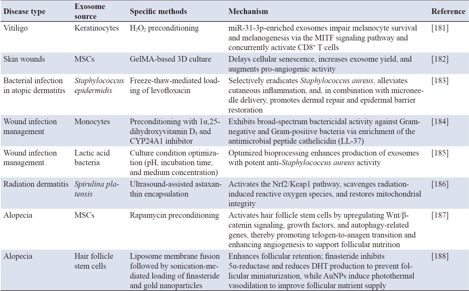
So far, we have focused on inflammation, autoimmunity, fibrosis, wound healing, and cancer. However, the same logic extends to other skin conditions that do not respond well to conventional treatments—pigmentary disorders, physical trauma, and cutaneous appendage diseases. Representative strategies are summarized in Table 2, ranging from melanin regulation to hair follicle regeneration.
Engineered exosomes represent a promising new tool for dermatology. Modifications can be introduced in terms of cargo, membrane, and production process. These rational designs translate into precise targeting, stronger therapeutic effects, and greater flexibility, enabling engineered exosomes to perform functions beyond those of natural vesicles. This implies that a single platform could perform multiple therapeutic functions. Due to this synergistic advantage, engineered exosomes have been applied to distinct types of skin diseases, including inflammatory disorders, fibrosis, impaired wound healing, and skin tumors.
Most studies focus on how to enhance the performance of engineered exosomes. Although a range of surface engineering strategies is available, it is essential to verify that these modifications do not alter native protein conformations or compromise the integrity of the lipid bilayer. This is a particular concern with chemical conjugation, as residual coupling reagents may persist and contribute to adverse reactions. Cargo loading presents a comparable constraint. In practice, many researchers apply mechanical treatments to exosomes, yet capacity and stability impose an upper limit on the number of molecules a single vesicle can accommodate. Liposome-exosome fusion is often used to increase drug loading, but it also blurs the boundary between exosomes and biomimetic vesicles. When typical markers such as CD63 and CD81 are weakened or lost, these hybrid vesicles may be better described as exosome mimetics rather than conventional exosomes. Protein nanoparticle co-assembly supraparticles offer a new strategy for enhancing payload capacity. Their compact size favors subsequent packaging into exosomes before secretion [189].
Vesicles derived from different sources possess distinct advantages, and those secreted by probiotics or beneficial skin commensals have attracted increasing interest as potential therapeutic agents for dermatologic intervention [183, 190]. Nevertheless, extraction and purification methods for microbial exosomes are still technically demanding. For scalable manufacturing, low-cost and readily accessible feedstocks, like plants and biofluids, are preferred. Milk exosome production, in particular, has reached a relatively mature stage of industrial development [191]. Regardless of scale, batch‑to‑batch consistency remains a critical requirement. Minor variations can lead to differences in the final product, so processing steps should ideally be carried out within an integrated, continuous workflow. Critical quality attributes and process parameters are central to regulatory approval. No universally accepted standard has yet been established for defining exosome purity, whether by particle number, protein content, or surface markers. To date, no drug regulatory agency has issued dedicated technical guidelines specific to exosomes, and classification varies across jurisdictions [192]. The core debate lies in whether exosomes should be classified as drugs or biologics. Xu et al. argued for positioning exosomes within the framework of advanced therapy medicinal products [193]. They also advised implementing refined oversight that distinguishes native from engineered exosomes. Engineered exosomes have opened a new avenue for the treatment of refractory skin diseases. Future progress will depend on closer coordination among design, manufacturing, quality control, and clinical translation, along with genuine interdisciplinary collaboration.
Author contributions
Jiayi Wang and Junkun Qu performed the image processing and wrote the manuscript. Don Green helped with language polishing of the manuscript. Yangyang Zhang and Yuling Chen contributed significantly to the analysis and manuscript editing. Yongtai Zhang contributed to the analysis through constructive discussions. Teng Guo and Nianping Feng performed the manuscript review, project administration, and resource acquisition.
Funding
This work was supported by the National Natural Science Foundation of China (82574706) and the Technology Development Project of Shanghai University of Traditional Chinese Medicine (25KJRH13).
Data availability
No new data were created or analyzed in this study.
Ethics approval and consent to participate
Not applicable.
Consent for publication
All authors have read the manuscript and agreed to its publication.
Competing interests
All authors declare that there are no competing interests.
Acknowledgements
Not applicable.
[1] Burden of 375 diseases and injuries, risk-attributable burden of 88 risk factors, and healthy life expectancy in 204 countries and territories, including 660 subnational locations, 1990-2023: A systematic analysis for the global burden of disease study 2023. Lancet. 2025 Oct 18;406(10513):1873-1922. https://doi.org/10.1016/s0140-6736(25)01637-x
[2] Cao F, Tian Z, Zhang H, Yu Q, Guo J. Dynamic evolution of the global burden of chronic urticaria: A comprehensive trend analysis from 1990 to 2021 and projections to 2050. Int J Surg. 2025 Dec 1;111(12):9471-9481. https://doi.org/10.1097/js9.0000000000003139
[3] Facheris P, Jeffery J, Del Duca E, Guttman-Yassky E. The translational revolution in atopic dermatitis: The paradigm shift from pathogenesis to treatment. Cell Mol Immunol. 2023 May;20(5):448-474. https://doi.org/10.1038/s41423-023-00992-4
[4] Nast A, Smith C, Spuls PI, Avila Valle G, Bata-Csörgö Z, Boonen H, et al. Euroguiderm guideline on the systemic treatment of psoriasis vulgaris - Part 1: Treatment and monitoring recommendations. J Eur Acad Dermatol Venereol. 2020 Nov;34(11):2461-2498. https://doi.org/10.1111/jdv.16915
[5] Cabrera-Perez JS, Carey VJ, Odejide OO, Singh S, Kupper TS, Pillai SS, et al. Integrative epidemiology and immunotranscriptomics uncover a risk and potential mechanism for cutaneous lymphoma unmasking or progression with dupilumab therapy. J Allergy Clin Immunol. 2025 May;155(5):1584-1594. https://doi.org/10.1016/j.jaci.2024.10.028
[6] van Niel G, D’Angelo G, Raposo G. Shedding light on the cell biology of extracellular vesicles. Nat Rev Mol Cell Biol. 2018 Apr;19(4):213-228. https://doi.org/10.1038/nrm.2017.125
[7] Kalluri R, LeBleu VS. The biology, function, and biomedical applications of exosomes. Science. 2020 Feb 7;367(6478):eaau6977. https://doi.org/10.1126/science.aau6977
[8] Marcianti A, Spampinato E, Nava S, Stella GM, Perego P, Pogliani S, et al. Extracellular vesicles isolated from adipose tissue-derived mesenchymal stromal cells as carriers for paclitaxel delivery. Stem Cell Res Ther. 2025 Jun 15;16(1):307. https://doi.org/10.1186/s13287-025-04435-x
[9] Witwer KW, Wolfram J. Extracellular vesicles versus synthetic nanoparticles for drug delivery. Nat Rev Mater. 2021 Feb;6(2):103-106. https://doi.org/10.1038/s41578-020-00277-6
[10] Kim J, Gao C, Guo P, Sheng J, Wang J. A novel approach to alleviate acetaminophen-induced hepatotoxicity with hybrid balloon flower root-derived exosome-like nanoparticles (BDEs) with silymarin via inhibition of hepatocyte MAPK pathway and apoptosis. Cell Commun Signal. 2024 Jun 18;22(1):334. https://doi.org/10.1186/s12964-024-01700-z
[11] Ding J, Chen M, Wu L, Shu G, Fang S, Li Z, et al. Mesenchymal stem cell-derived extracellular vesicles in skin wound healing: Roles, opportunities and challenges. Mil Med Res. 2023 Aug 17;10(1):36. https://doi.org/10.1186/s40779-023-00472-w
[12] Tutuianu R, Rosca AM, Iacomi DM, Simionescu M, Titorencu I. Human mesenchymal stromal cell-derived exosomes promote in vitro wound healing by modulating the biological properties of skin keratinocytes and fibroblasts and stimulating angiogenesis. Int J Mol Sci. 2021 Jun 9;22(12):6239. https://doi.org/10.3390/ijms22126239
[13] Zhou Y, Zhang X, Lu S, Zhang N, Zhang H, Zhang J, et al. Human adipose-derived mesenchymal stem cells-derived exosomes encapsulated in pluronic F127 hydrogel promote wound healing and regeneration. Stem Cell Res Ther. 2022 Aug 8;13(1):407. https://doi.org/10.1186/s13287-022-02980-3
[14] Yang Y, Huang Y, Yang J, Hu Z, Wu S, Yuan Q, et al. Umbilical cord mesenchymal stem cell-derived exosomes promote wound healing and skin regeneration via the regulation of inflammation and angiogenesis. Front Bioeng Biotechnol. 2025 Nov;13:1641709. https://doi.org/10.3389/fbioe.2025.1641709
[15] Shao J, Xie Z, Ye Z, Chen G, Wang Y, Zhang L, et al. Human umbilical cord mesenchymal stem cell therapy for atopic dermatitis through inhibition of neutrophil chemotaxis. Stem Cell Res Ther. 2025 May 14;16(1):243. https://doi.org/10.1186/s13287-025-04349-8
[16] Wang S, Zhang Y, Zeng Y, Luo X, Chen J, Deng Q, et al. Plant-derived vesicle-like nanoparticles for immunomodulation: Mechanisms and applications. Bioact Mater. 2026 Jan;55:171-204. https://doi.org/10.1016/j.bioactmat.2025.09.024
[17] Wang Z, Yuan J, Xu Y, Shi N, Lin L, Wang R, et al. Olea europaea leaf exosome-like nanovesicles encapsulated in a hyaluronic acid / tannic acid hydrogel dressing with dual “defense-repair” effects for treating skin photoaging. Mater Today Bio. 2024 Jun;26:101103. https://doi.org/10.1016/j.mtbio.2024.101103
[18] Kim HI, Park J, Zhu Y, Wang X, Han Y, Zhang D. Recent advances in extracellular vesicles for therapeutic cargo delivery. Exp Mol Med. 2024 Apr;56(4):836-849. https://doi.org/10.1038/s12276-024-01201-6
[19] Pascucci L, Coccè V, Bonomi A, Ami D, Ceccarelli P, Ciusani E, et al. Paclitaxel is incorporated by mesenchymal stromal cells and released in exosomes that inhibit in vitro tumor growth: A new approach for drug delivery. J Control Release. 2014 Oct 28;192:262-270. https://doi.org/10.1016/j.jconrel.2014.07.042
[20] Alvarez-Erviti L, Seow Y, Yin H, Betts C, Lakhal S, Wood MJ. Delivery of siRNA to the mouse brain by systemic injection of targeted exosomes. Nat Biotechnol. 2011 Apr;29(4):341-345. https://doi.org/10.1038/nbt.1807
[21] Liu Q, Li D, Pan X, Liang Y. Targeted therapy using engineered extracellular vesicles: Principles and strategies for membrane modification. J Nanobiotechnology. 2023 Sep 16;21(1):334. https://doi.org/10.1186/s12951-023-02081-0
[22] Chen Z, Xiong M, Tian J, Song D, Duan S, Zhang L. Encapsulation and assessment of therapeutic cargo in engineered exosomes: A systematic review. J Nanobiotechnology. 2024 Jan 3;22(1):18. https://doi.org/10.1186/s12951-023-02259-6
[23] Huang J, Chen H, Li N, Liu P, Yang J, Zhao Y. Emerging technologies towards extracellular vesicles large-scale production. Bioact Mater. 2025 Oct;52:338-365. https://doi.org/10.1016/j.bioactmat.2025.06.005
[24] Qiu T, Liu Z, Zhang H, Jia J, Shao X, Wang S, et al. Engineered exosomes with enriched miR-23b suppress the progression of periodontitis by reprogramming macrophages. Colloids Surf B Biointerfaces. 2025 Nov;255:114947. https://doi.org/10.1016/j.colsurfb.2025.114947
[25] Yueh PF, Chiang IT, Weng YS, Liu YC, Wong RCB, Chen CY, et al. Innovative dual-gene delivery platform using miR-124 and PD-1 via umbilical cord mesenchymal stem cells and exosome for glioblastoma therapy. J Exp Clin Cancer Res. 2025 Mar 25;44(1):107. https://doi.org/10.1186/s13046-025-03336-4
[26] Peruzzi JA, Gunnels TF, Edelstein HI, Lu P, Baker D, Leonard JN, et al. Enhancing extracellular vesicle cargo loading and functional delivery by engineering protein-lipid interactions. Nat Commun. 2024 Jul 4;15(1):5618. https://doi.org/10.1038/s41467-024-49678-z
[27] Zickler AM, Liang X, Gupta D, Mamand DR, De Luca M, Corso G, et al. Novel endogenous engineering platform for robust loading and delivery of functional mRNA by extracellular vesicles. Adv Sci (Weinh). 2024 Nov;11(42):e2407619. https://doi.org/10.1002/advs.202407619
[28] Liang X, Gupta D, Xie J, Van Wonterghem E, Van Hoecke L, Hean J, et al. Engineering of extracellular vesicles for efficient intracellular delivery of multimodal therapeutics including genome editors. Nat Commun. 2025 Apr 29;16(1):4028. https://doi.org/10.1038/s41467-025-59377-y
[29] Elsharkasy OM, Hegeman CV, Driedonks TAP, Liang X, Lansweers I, Cotugno OL, et al. A modular strategy for extracellular vesicle-mediated CRISPR-Cas9 delivery through aptamer-based loading and UV-activated cargo release. Nat Commun. 2025 Nov 21;16(1):10309. https://doi.org/10.1038/s41467-025-65995-3
[30] Hegeman CV, Elsharkasy OM, Driedonks TAP, Friesen KRJ, Vader P, de Jong OG. Modulating binding affinity of aptamer-based loading constructs enhances extracellular vesicle-mediated CRISPR/Cas9 delivery. J Control Release. 2025 Aug 10;384:113853. https://doi.org/10.1016/j.jconrel.2025.113853
[31] Cao X, Wu Y, Shen Y, Xu Z, Zhang X, Fan Z, et al. Extracellular vesicles from hypoxia preconditioned bone marrow mesenchymal stem cell improve peri-implant osteogenesis under type 2 diabetes condition. J Control Release. 2025 Dec 10;388(Pt 1):114276. https://doi.org/10.1016/j.jconrel.2025.114276
[32] Toghiani R, Azimian Zavareh V, Najafi H, Mirian M, Azarpira N, Abolmaali SS, et al. Hypoxia-preconditioned WJ-MSC spheroid-derived exosomes delivering miR-210 for renal cell restoration in hypoxia-reoxygenation injury. Stem Cell Res Ther. 2024 Jul 30;15(1):240. https://doi.org/10.1186/s13287-024-03845-7
[33] Kim JY, Rhim WK, Woo J, Cha SG, Park CG, Han DK. The upregulation of regenerative activity for extracellular vesicles with melatonin modulation in chemically defined media. Int J Mol Sci. 2022 Dec 1;23(23):15089. https://doi.org/10.3390/ijms232315089
[34] Wu J, Wu J, Xiang W, Gong Y, Feng D, Fang S, et al. Engineering exosomes derived from TNF-α preconditioned IPFP-MSCs enhance both yield and therapeutic efficacy for osteoarthritis. J Nanobiotechnology. 2024 Sep 11;22(1):555. https://doi.org/10.1186/s12951-024-02795-9
[35] Yao W, Zhou J, Tang C, Zhang J, Chen Z, Li Y, et al. Hydrogel microneedle patches loaded with stem cell mitochondria-enriched microvesicles boost the chronic wound healing. ACS Nano. 2024 Oct 1;18(39):26733-26750. https://doi.org/10.1021/acsnano.4c06921
[36] Liu H, Liu Y, Peng S, Zhou L, McClements DJ, Fang S, et al. Colonic delivery and controlled release of curcumin encapsulated within plant-based extracellular vesicles loaded into hydrogel beads. Food Res Int. 2025 Feb;202:115540. https://doi.org/10.1016/j.foodres.2024.115540
[37] Schifano E, Vari F, Buccini L, Karimova M, Syman K, Varnadyan D, et al. A novel scalable method for the production of rennet-treated milk-derived extracellular vesicles for improved curcumin oral delivery. J Nanobiotechnology. 2025 Oct 10;23(1):656. https://doi.org/10.1186/s12951-025-03724-0
[38] Song L, Tang Y, Qu Y, Yun Y, Zhang R, Wang C, et al. Exosomal delivery of rapamycin modulates blood-brain barrier penetration and VEGF axis in glioblastoma. J Control Release. 2025 May 10;381:113605. https://doi.org/10.1016/j.jconrel.2025.113605
[39] Jiang Q, Sun Y, Si X, Cui H, Li J, Bao Y, et al. Anthocyanin-loaded milk-derived extracellular vesicles nano-delivery system: Stability, mucus layer penetration, and pro-oxidant effect on HepG2 cells. Food Chem. 2024 Nov 15;458:140152. https://doi.org/10.1016/j.foodchem.2024.140152
[40] Chung J, Kim KH, An SH, Bae D, Kim HK, Choi Y, et al. A single extracellular vesicle-based platform supporting both RBD protein and mRNA vaccination against SARS-CoV-2. J Control Release. 2026 Feb 10;390:114515. https://doi.org/10.1016/j.jconrel.2025.114515
[41] Pan X, Huang P, Ali SS, Renslo B, Greenberg Z, Erwin N, et al. Extracellular vesicle-mediated gene editing for the treatment of nonsyndromic progressive hearing loss in adult mice. Sci Transl Med. 2025 Nov 12;17(824):eadn3993. https://doi.org/10.1126/scitranslmed.adn3993
[42] Kim YM, Kim H, Park SC, Lee M, Jang MK. Targeted drug delivery of cancer cell-derived extracellular vesicles decorated with a VEGFR-binding peptide. Colloids Surf B Biointerfaces. 2025 Aug;252:114661. https://doi.org/10.1016/j.colsurfb.2025.114661
[43] Xiao P, Wang H, Liu H, Yuan H, Guo C, Feng Y, et al. Milk exosome-liposome hybrid vesicles with self-adapting surface properties overcome the sequential absorption barriers for oral delivery of peptides. ACS Nano. 2024 Aug 13;18(32):21091-21111. https://doi.org/10.1021/acsnano.4c02560
[44] Wang X, Munson MJ, Friis K, Marzeda A, Silva AM, Kohl F, et al. Hybrid extracellular vesicles for efficient loading and functional delivery of mRNA. J Extracell Vesicles. 2025 Dec;14(12):e70201. https://doi.org/10.1002/jev2.70201
[45] Son G, Song J, Park JC, Kim HN, Kim H. Fusogenic lipid nanoparticles for rapid delivery of large therapeutic molecules to exosomes. Nat Commun. 2025 May 23;16(1):4799. https://doi.org/10.1038/s41467-025-59489-5
[46] Abdel-Bar HM, Tandiono S, Liam-Or R, Cheung CCL, Hassuneh OWM, Lyu Q, et al. Optimizing exosome lipid hybrid nanoparticles for enhanced siRNA delivery and improved therapeutic anticancer efficacy in vivo. ACS Nano. 2025 Dec 23;19(50):42658-42674. https://doi.org/10.1021/acsnano.5c16991
[47] Yang Z, Shi J, Xie J, Wang Y, Sun J, Liu T, et al. Large-scale generation of functional mRNA-encapsulating exosomes via cellular nanoporation. Nat Biomed Eng. 2020 Jan;4(1):69-83. https://doi.org/10.1038/s41551-019-0485-1
[48] You Y, Tian Y, Yang Z, Shi J, Kwak KJ, Tong Y, et al. Intradermally delivered mRNA-encapsulating extracellular vesicles for collagen-replacement therapy. Nat Biomed Eng. 2023 Jul;7(7):887-900. https://doi.org/10.1038/s41551-022-00989-w
[49] You Y, Tian Y, Guo R, Shi J, Kwak KJ, Tong Y, et al. Extracellular vesicle-mediated VEGF-A mRNA delivery rescues ischaemic injury with low immunogenicity. Eur Heart J. 2025 May 2;46(17):1662-1676. https://doi.org/10.1093/eurheartj/ehae883
[50] Sheng Y, Huang Z, Zhang T, Qian F, Zhu Y, Dong Z, et al. Living cell nanoporation and exosomal RNA analysis platform for real-time assessment of cellular therapies. J Am Chem Soc. 2022 Jun 1;144(21):9443-9450. https://doi.org/10.1021/jacs.2c02268
[51] Ramon J, Pinheiro C, Vandendriessche C, Lozano-Andrés E, De Keersmaecker H, Punj D, et al. Pre-formation loading of extracellular vesicles with exogenous molecules using photoporation. J Nanobiotechnology. 2025 Aug 8;23(1):556. https://doi.org/10.1186/s12951-025-03640-3
[52] Li C, Wen Y, Wang J, Li L, He Y, Cheng Y, et al. Human mesenchymal stem cell-derived exosomes as engineering vehicles of daunorubicin for targeted c-Mpl+ AML therapy. Int J Nanomedicine. 2025 Apr;20:5267-5289. https://doi.org/10.2147/ijn.S511713
[53] Liu X, Liu X, Luo X, Zhu M, Liu N, Li J, et al. Synergistic strategies for glioblastoma treatment: Crispr-based multigene editing combined with immune checkpoint blockade. J Nanobiotechnology. 2025 Feb 7;23(1):94. https://doi.org/10.1186/s12951-025-03112-8
[54] Zhu Z, Zhai Y, Hao Y, Wang Q, Han F, Zheng W, et al. Specific anti-glioma targeted-delivery strategy of engineered small extracellular vesicles dual-functionalised by Angiopep-2 and TAT peptides. J Extracell Vesicles. 2022 Aug;11(8):e12255. https://doi.org/10.1002/jev2.12255
[55] Xu F, Fei Z, Dai H, Xu J, Fan Q, Shen S, et al. Mesenchymal stem cell-derived extracellular vesicles with high PD-L1 expression for autoimmune diseases treatment. Adv Mater. 2022 Jan;34(1):e2106265. https://doi.org/10.1002/adma.202106265
[56] Zhou G, Gu Y, Zhang M, Ding J, Lu G, Hua K, et al. Identification of genetically engineered strategies to manipulate nanoplatforms presenting immunotherapeutic ligands for alleviating primary ovarian insufficiency progression. Cell Commun Signal. 2025 May 28;23(1):246. https://doi.org/10.1186/s12964-025-02226-8
[57] Cheng L, Zhang X, Tang J, Lv Q, Liu J. Gene-engineered exosomes-thermosensitive liposomes hybrid nanovesicles by the blockade of CD47 signal for combined photothermal therapy and cancer immunotherapy. Biomaterials. 2021 Aug;275:120964. https://doi.org/10.1016/j.biomaterials.2021.120964
[58] Du W, Chen C, Liu Y, Quan H, Xu M, Liu J, et al. A combined “eat me/don’t eat me” strategy based on exosome for acute liver injury treatment. Cell Rep Med. 2025 Apr 15;6(4):102033. https://doi.org/10.1016/j.xcrm.2025.102033
[59] Zhao H, Li Z, Liu D, Zhang J, You Z, Shao Y, et al. PlexinA1 (PLXNA1) as a novel scaffold protein for the engineering of extracellular vesicles. J Extracell Vesicles. 2024 Nov;13(11):e70012. https://doi.org/10.1002/jev2.70012
[60] Zhao N, Guo Z, Bai S, Gao Y, Lei J, Li P, et al. Endothelial cell-targeting aptamer-empowered exosomes accelerate wound healing by promoting specialized angiogenesis in type 1 diabetic mice. Stem Cell Res Ther. 2025 Dec 7;17(1):23. https://doi.org/10.1186/s13287-025-04846-w
[61] Li Z, Yang L, Wang H, Binzel DW, Williams TM, Guo P. Non-small-cell lung cancer regression by siRNA delivered through exosomes that display EGFR RNA aptamer. Nucleic Acid Ther. 2021 Oct;31(5):364-374. https://doi.org/10.1089/nat.2021.0002
[62] Zheng W, Schürz M, Wiklander RJ, Gustafsson O, Gupta D, Slovak R, et al. Surface display of functional moieties on extracellular vesicles using lipid anchors. J Control Release. 2023 May;357:630-640. https://doi.org/10.1016/j.jconrel.2023.04.033
[63] Hosseini NF, Amini R, Ramezani M, Saidijam M, Hashemi SM, Najafi R. AS1411 aptamer-functionalized exosomes in the targeted delivery of doxorubicin in fighting colorectal cancer. Biomed Pharmacother. 2022 Nov;155:113690. https://doi.org/10.1016/j.biopha.2022.113690
[64] Tian J, Wang Z, Tang Z, Zhu H, Sun L. Engineered exosomes conferred with ROS-regulation and immuno-suppression for ameliorating lupus nephritis. J Nanobiotechnology. 2025 Oct 10;23(1):639. https://doi.org/10.1186/s12951-025-03731-1
[65] Chen C, Pan X, Sun M, Wang J, Su X, Zhang T, et al. Phospholipid-anchored ligand conjugation on extracellular vesicles for enhanced cancer targeting. Small. 2024 Sep;20(37):e2310712. https://doi.org/10.1002/smll.202310712
[66] Agatemor C, Buettner MJ, Ariss R, Muthiah K, Saeui CT, Yarema KJ. Exploiting metabolic glycoengineering to advance healthcare. Nat Rev Chem. 2019 Oct;3(10):605-620. https://doi.org/10.1038/s41570-019-0126-y
[67] Chen P, Wang R, Fang S. Metabolic glycoengineered exosome-A2M nanoplatform reprograms macrophage polarization and orchestrates bone regeneration in ONFH. Cell Death Discov. 2025 Nov 7;11(1):510. https://doi.org/10.1038/s41420-025-02690-8
[68] Tong Y, Sun J, Jiang X, Jia X, Xiao H, Wang H, et al. A study on the production of extracellular vesicles derived from novel immortalized human placental mesenchymal stromal cells. Sci Rep. 2025 Jan 28;15(1):3568. https://doi.org/10.1038/s41598-025-87371-3
[69] Tong Y, Sun J, Jiang X, Jia X, Jia Y, Wang H, et al. Stable production of immortalized hPC-MSC-derived extracellular vesicles: Integrated cargo analysis and functional validation. Stem Cell Res Ther. 2025 Nov 5;16(1):619. https://doi.org/10.1186/s13287-025-04743-2
[70] Gong S, Li N, Peng Q, Wang F, Du R, Zhang B, et al. A scalable platform for EPSC-induced MSC extracellular vesicles with therapeutic potential. Stem Cell Res Ther. 2025 Aug 5;16(1):426. https://doi.org/10.1186/s13287-025-04507-y
[71] Uman S, Worthington K, Dominic J, Atluri P, Burdick JA. Comparison of technologies for manufacturing extracellular vesicles for therapeutic applications. Trends Biotechnol. 2026 Apr;44(4):1079-1099. https://doi.org/10.1016/j.tibtech.2025.11.005
[72] Tan J, Hu Y, Zheng L, Zheng Z, Fu M, Peng H, et al. Microbead encapsulation strategy for efficient production of extracellular vesicles derived from human mesenchymal stem cells. J Extracell Vesicles. 2025 Apr;14(4):e70053. https://doi.org/10.1002/jev2.70053
[73] Ding Y, Fu Y, Sun M, Li Y, Li A, Li Y. Microgravity-driven Rab27B activation amplifies mesenchymal stem cell-derived extracellular vesicle production and functions. Stem Cell Res Ther. 2025 Oct 29;16(1):590. https://doi.org/10.1186/s13287-025-04711-w
[74] Thouvenot E, Charnay L, Burshtein N, Guigner JM, Dec L, Loew D, et al. High-yield bioproduction of extracellular vesicles from stem cell spheroids via millifluidic vortex transport. Adv Mater. 2025 May;37(20):e2412498. https://doi.org/10.1002/adma.202412498
[75] Chintapula U, Liu S, Castillo AFD, Lim J, Roh Y, Mageswaran SK, et al. Droplet squeeze microfluidic platform for generating extracellular vesicle hybrids for drug delivery. Small. 2025 Sep;21(37):e2503807. https://doi.org/10.1002/smll.202503807
[76] Xiao L, Wang J, Yu M, Chen T, You J, Li Q, et al. Emerging nutritional potential of edible-medicinal homologous coix lacryma-jobi seed-derived exosomes for treatment of ulcerative colitis. Int J Biol Macromol. 2025 Dec;332(Pt 2):148799. https://doi.org/10.1016/j.ijbiomac.2025.148799
[77] Del Saz-Lara A, López de Las Hazas MC, Tomé-Carneiro J, Mazarío-Gárgoles C, Gil-Zamorano J, Balaguer L, et al. Milk and cheesemaking whey as a sustainable source of extracellular vesicles: Exploring large-scale isolation methods with industrial and potential therapeutic applications. Pharmacol Res. 2025 Oct;220:107925. https://doi.org/10.1016/j.phrs.2025.107925
[78] Cao Y, Qin Y, Cheng Q, Zhong J, Han B, Li Y. Bifunctional nanomaterial enabled high-specific isolation of urinary exosomes for cervical cancer metabolomics analysis and biomarker discovery. Talanta. 2025 Apr 1;285:127280. https://doi.org/10.1016/j.talanta.2024.127280
[79] Weerakkody JS, Tseng T, Topper M, Thoduvayil S, Radhakrishnan A, Pincet F, et al. Photosensitive nanoprobes for rapid isolation and size-specific enrichment of synthetic and extracellular vesicle subpopulations. Adv Funct Mater. 2024 Aug 22;34(34):2400390. https://doi.org/10.1002/adfm.202400390
[80] Marttila H, Saari H, Laitinen S, Poranen MM, Oksanen HM. Separation of platelet extracellular vesicles from lipoproteins by monolithic anion-exchange chromatography. J Chromatogr A. 2025 Nov 22;1763:466416. https://doi.org/10.1016/j.chroma.2025.466416
[81] Woo HK, Cho YK, Lee CY, Lee H, Castro CM, Lee H. Characterization and modulation of surface charges to enhance extracellular vesicle isolation in plasma. Theranostics. 2022 Jan;12(5):1988-1998. https://doi.org/10.7150/thno.69094
[82] Chen Y, Zhu Q, Cheng L, Wang Y, Li M, Yang Q, et al. Exosome detection via the ultrafast-isolation system: Exodus. Nat Methods. 2021 Feb;18(2):212-218. https://doi.org/10.1038/s41592-020-01034-x
[83] Wang Z, Qin Z, Wang J, Xu X, Zhang M, Liang Y, et al. Engineering extracellular vesicles with macrophage membrane fusion for ameliorating imiquimod-induced psoriatic skin inflammation. J Dermatolog Treat. 2023 Dec;34(1):2220445. https://doi.org/10.1080/09546634.2023.2220445
[84] Zhang H, Sun M, Chen J, Wang S, Liu J, Yang H, et al. Immunological conversion of triple-negative breast cancer by engineered extracellular vesicle-mediated pyroptosis. J Control Release. 2026 Jan 10;389:114493. https://doi.org/10.1016/j.jconrel.2025.114493
[85] Wu X, Tao W, Lan Z, Tian Y, Zhong Z, Wang J, et al. pH-responsive engineered exosomes enhance endogenous hyaluronan production by reprogramming chondrocytes for cartilage repair. Adv Healthc Mater. 2025 Apr;14(10):e2405126. https://doi.org/10.1002/adhm.202405126
[86] Abraham AM, Wiemann S, Ambreen G, Zhou J, Engelhardt K, Brüßler J, et al. Cucumber-derived exosome-like vesicles and plantcrystals for improved dermal drug delivery. Pharmaceutics. 2022 Feb 22;14(3):476. https://doi.org/10.3390/pharmaceutics14030476
[87] Gu Y, Bian Q, Zhou Y, Huang Q, Gao J. Hair follicle-targeting drug delivery strategies for the management of hair follicle-associated disorders. Asian J Pharm Sci. 2022 May;17(3):333-352. https://doi.org/10.1016/j.ajps.2022.04.003
[88] Ma Y, Dong J, Li M, Du X, Yan Z, Tian W. An antimicrobial microneedle patch promotes functional healing of infected wounds through controlled release of adipose tissue-derived apoptotic vesicles. J Nanobiotechnology. 2024 Sep 20;22(1):579. https://doi.org/10.1186/s12951-024-02845-2
[89] Li Y, Qian L, Liu F, Xu S, Zhou L, Wei C, et al. Light-controlled small extracellular vesicle-based spherical nucleic acid nanomotor for enhanced transdermal delivery against skin aging. Nano Lett. 2025 Jun 4;25(22):9098-9109. https://doi.org/10.1021/acs.nanolett.5c01747
[90] Obuchi W, Zargani-Piccardi A, Leandro K, Rufino-Ramos D, Di Lanni E, Frederick DM, et al. Engineering of CD63 enables selective extracellular vesicle cargo loading and enhanced payload delivery. J Extracell Vesicles. 2025 Jun;14(6):e70094. https://doi.org/10.1002/jev2.70094
[91] Lu Y, Guo Y, Wang L, Nakamura Y, Qi H. Royal jelly exosome-based nanocarriers enhance the stability and bioactivity of polyphenols from ascophyllum nodosum for melanin regulatio. Food Res Int. 2025 Dec;221(Pt 4):117495. https://doi.org/10.1016/j.foodres.2025.117495
[92] Cheng Y, Zeng Q, Han Q, Xia W. Effect of pH, temperature and freezing-thawing on quantity changes and cellular uptake of exosomes. Protein Cell. 2019 Apr;10(4):295-299. https://doi.org/10.1007/s13238-018-0529-4
[93] Kim J, Hwang YH, Nam GH, Kim IS. Breaking barriers: Engineering extracellular vesicles for enhanced endosomal escape and therapeutic delivery. J Control Release. 2026 Jan 10;389:114462. https://doi.org/10.1016/j.jconrel.2025.114462
[94] Zhang J, Li J, Zheng Y, Zhu C, Wu Z, Wang T, et al. Lipid metabolism and lipid signaling in extracellular vesicles ontogeny: From biogenesis to functional execution. J Nanobiotechnology. 2025 Oct 14;23(1):672. https://doi.org/10.1186/s12951-025-03769-1
[95] Qin Y, Chen X, Bao L, Ren L, Dou G, Lian J, et al. Lipid metabolism of apoptotic vesicles accelerates cutaneous wound healing by modulating macrophage function. J Nanobiotechnology. 2025 Feb 12;23(1):106. https://doi.org/10.1186/s12951-025-03194-4
[96] Osteikoetxea X, Silva A, Lázaro-Ibáñez E, Salmond N, Shatnyeva O, Stein J, et al. Engineered Cas9 extracellular vesicles as a novel gene editing tool. J Extracell Vesicles. 2022 May;11(5):e12225. https://doi.org/10.1002/jev2.12225
[97] Shi A, Li J, Qiu X, Sabbah M, Boroumand S, Huang TC, et al. TGF-β loaded exosome enhances ischemic wound healing in vitro and in vivo. Theranostics. 2021 Apr 30;11(13):6616-6631. https://doi.org/10.7150/thno.57701
[98] Jia S, Yang Y, Liu J, Wang Z, Bai L. PPARγ controls ESCRT-dependent fibroblast-like synoviocyte exosome biogenesis and alleviates chondrocyte osteoarthritis mediated by exosomal ANXA1. J Orthop Translat. 2025 Jul;53:187-205. https://doi.org/10.1016/j.jot.2025.06.008
[99] Chen Y, Yin W, Liu Z, Lu G, Zhang X, Yang J, et al. Exosomes derived from fibroblasts enhance skin wound angiogenesis by regulating HIF-1α/VEGF/VEGFR pathway. Burns Trauma. 2025 May;13:tkae071. https://doi.org/10.1093/burnst/tkae071
[100] Yan L, Fan D, Yang J, Wang J, Hu X, Zhang X, et al. Fibroblast exosomes promote wound healing and improve the quality of healed skin via miR-29a-3p-mediated KEAP1/Nrf2 pathway activation. Burns Trauma. 2025 May;13:tkaf035. https://doi.org/10.1093/burnst/tkaf035
[101] Liu B, Li X, Yu H, Shi X, Zhou Y, Alvarez S, et al. Therapeutic potential of garlic chive-derived vesicle-like nanoparticles in NLRP3 inflammasome-mediated inflammatory diseases. Theranostics. 2021 Sep 7;11(19):9311-9330. https://doi.org/10.7150/thno.60265
[102] Niu L, Ou Q, Ren Q, Li Z, Chen H, Lei F, et al. Engineered foxp1(high) exosomes ameliorates systemic lupus erythematosus. Adv Sci (Weinh). 2025 Oct;12(37):e15712. https://doi.org/10.1002/advs.202415712
[103] Wu M, Tu J, Huang J, Wen H, Zeng Y, Lu Y. Exosomal IRF1-loaded rat adipose-derived stem cell sheet contributes to wound healing in the diabetic foot ulcers. Mol Med. 2023 Apr 25;29(1):60. https://doi.org/10.1186/s10020-023-00617-6
[104] Gao Y, Liu J, Wu M, Zhang Y, Wang M, Lyu Q, et al. Photosensitive hybrid γδ-T exosomes for targeted cancer photoimmunotherapy. ACS Nano. 2025 Feb 4;19(4):4251-4268. https://doi.org/10.1021/acsnano.4c11024
[105] Ma W, Che J, Chen W, Wang D, Zhang H, Zhao Y. Dexamethasone-integrated mesenchymal stem cells for systemic lupus erythematosus treatment via multiple immunomodulatory mechanisms. ACS Nano. 2024 May 21;18(20):13249-13265. https://doi.org/10.1021/acsnano.4c02420
[106] Ou M, Cao J, Luo R, Zhu B, Miao R, Yu L, et al. Drug-loaded microneedle patches containing regulatory T cell-derived exosomes for psoriasis treatment. Acta Biomater. 2025 May 15;198:452-466. https://doi.org/10.1016/j.actbio.2025.04.015
[107] Han B, Fang W, Yang Z, Wang Y, Zhao S, Hoang BX, et al. Enhancement of chondrogenic markers by exosomes derived from cultured human synovial fluid-derived cells: A comparative analysis of 2D and 3D conditions. Biomedicines. 2023 Nov 25;11(12):3145. https://doi.org/10.3390/biomedicines11123145
[108] Huang H, Xiao L, Fang L, Lei M, Liu Z, Gao S, et al. Static topographical cue combined with dynamic fluid stimulation enhances the macrophage extracellular vesicle yield and therapeutic potential for bone defects. ACS Nano. 2025 Mar 11;19(9):8667-8691. https://doi.org/10.1021/acsnano.4c15201
[109] Paganini C, Boyce H, Libort G, Arosio P. High-yield production of extracellular vesicle subpopulations with constant quality using batch-refeed cultures. Adv Healthc Mater. 2023 Mar;12(8):e2202232. https://doi.org/10.1002/adhm.202202232
[110] Kim JY, Rhim WK, Yoo YI, Kim DS, Ko KW, Heo Y, et al. Defined MSC exosome with high yield and purity to improve regenerative activity. J Tissue Eng. 2021 Jan-Dec;12:20417314211008626. https://doi.org/10.1177/20417314211008626
[111] Ghoreschi K, Balato A, Enerbäck C, Sabat R. Therapeutics targeting the IL-23 and IL-17 pathway in psoriasis. Lancet. 2021 Feb 20;397(10275):754-766. https://doi.org/10.1016/s0140-6736(21)00184-7
[112] Armstrong AW, Blauvelt A, Callis Duffin K, Huang YH, Savage LJ, Guo L, et al. Psoriasis. Nat Rev Dis Primers. 2025 Jun 26;11(1):45. https://doi.org/10.1038/s41572-025-00630-5
[113] Hong D, Xiong H, Lu S, Ma J, Shi Z. Metabolic regulation of the immune cell in psoriasis: Mechanisms and interventions. Curr Opin Immunol. 2025 Oct;96:102614. https://doi.org/10.1016/j.coi.2025.102614
[114] Cai J, Zhou X, Zhuang Y, Cui L, Ma R, Chen Y, et al. Reprogramming of fatty acid metabolism via PPARα-orchestrated FADS2 in keratinocytes modulates skin inflammation in psoriasis. Adv Sci (Weinh). 2025 Oct;12(40):e17049. https://doi.org/10.1002/advs.202417049
[115] Zhou Y, Zhou Y, Zhu Q, Guo W, Li C. Metabolic traits of T cells and the implications in autoimmune diseases. Autoimmun Rev. 2026 Feb;25(2):103989. https://doi.org/10.1016/j.autrev.2026.103989
[116] Gelfand JM. Psoriasis - more progress but more questions. N Engl J Med. 2024 Feb 8;390(6):561-562. https://doi.org/10.1056/NEJMe2314345
[117] Jairath V, Acosta Felquer ML, Cho RJ. IL-23 inhibition for chronic inflammatory disease. Lancet. 2024 Oct 26;404(10463):1679-1692. https://doi.org/10.1016/s0140-6736(24)01750-1
[118] Zhou X, Tang B, Huang Q, Yang S, Jiang Y, Xu L, et al. Engineered mesenchymal stem cell-derived extracellular vesicles scavenge self-antigens for psoriasis therapy via modulating metabolic and immunological disorders. Adv Sci (Weinh). 2025 Feb;12(6):e2410067. https://doi.org/10.1002/advs.202410067
[119] Liu L, Yang D, Ji J, Li G, Chen H, Yao Y, et al. Harnessing the immunomodulation of UV-exposed keratinocyte extracellular vesicles for inflammatory disorder treatment. Adv Sci (Weinh). 2025 Sep;12(36):e01517. https://doi.org/10.1002/advs.202501517
[120] Jiang X, Jiang Z, Huang S, Mao P, Zhang L, Wang M, et al. Ultraviolet B radiation-induced JPH203-loaded keratinocyte extracellular vesicles exert etiological interventions for psoriasis therapy. J Control Release. 2023 Oct;362:468-478. https://doi.org/10.1016/j.jconrel.2023.08.059
[121] Wolfson RL, Chantranupong L, Saxton RA, Shen K, Scaria SM, Cantor JR, et al. Sestrin2 is a leucine sensor for the mTORC1 pathway. Science. 2016 Jan 1;351(6268):43-48. https://doi.org/10.1126/science.aab2674
[122] Kishta MS, Abd-Rabou AA, Sarkissian GK, Elwakil AI, Elsabry DM, Zagzoug YM, et al. Exosome mediated delivery of Epigallocatechin 3 gallate as a novel approach to alleviate psoriasis symptoms through cytokine and apoptotic pathway modulation. Sci Rep. 2025 Aug 16;15(1):30013. https://doi.org/10.1038/s41598-025-10886-2
[123] Gao Y, Ma B, Jin R, Shao Y, Zhang L, Qi X, et al. Anhydroicaritin-loaded mesenchymal stem cell exosomes ameliorate psoriasis via ACSL4-mediated ferroptosis in mice. Commun Biol. 2026 Jan 23;9(1):306. https://doi.org/10.1038/s42003-026-09575-1
[124] Zhang W, Lin J, Shi P, Su D, Cheng X, Yi W, et al. Small extracellular vesicles derived from MSCs have immunomodulatory effects to enhance delivery of ASO-210 for psoriasis treatment. Front Cell Dev Biol. 2022 Mar;10:842813. https://doi.org/10.3389/fcell.2022.842813
[125] Liu N, Zhang J, Yin M, Liu H, Zhang X, Li J, et al. Inhibition of xCT suppresses the efficacy of anti-PD-1/L1 melanoma treatment through exosomal PD-L1-induced macrophage M2 polarization. Mol Ther. 2021 Jul 7;29(7):2321-2334. https://doi.org/10.1016/j.ymthe.2021.03.013
[126] Zhao Z, Zhang S, Yu R, Wang Z, Sun X, Zhang Z, et al. Optical microneedle-enhanced transdermal light scattering for in situ photothermal therapy targeting basal-layer psoriasis. ACS Appl Mater Interfaces. 2025 Apr 2;17(13):19446-19458. https://doi.org/10.1021/acsami.4c23014
[127] Kolkhir P, Akdis CA, Akdis M, Bachert C, Bieber T, Canonica GW, et al. Type 2 chronic inflammatory diseases: Targets, therapies and unmet needs. Nat Rev Drug Discov. 2023 Sep;22(9):743-767. https://doi.org/10.1038/s41573-023-00750-1
[128] Guttman-Yassky E, Renert-Yuval Y, Brunner PM. Atopic dermatitis. Lancet. 2025 Feb 15;405(10478):583-596. https://doi.org/10.1016/s0140-6736(24)02519-4
[129] Kubo A, Nagao K, Amagai M. Epidermal barrier dysfunction and cutaneous sensitization in atopic diseases. J Clin Invest. 2012 Feb;122(2):440-447. https://doi.org/10.1172/jci57416
[130] Palmer CN, Irvine AD, Terron-Kwiatkowski A, Zhao Y, Liao H, Lee SP, et al. Common loss-of-function variants of the epidermal barrier protein filaggrin are a major predisposing factor for atopic dermatitis. Nat Genet. 2006 Apr;38(4):441-446. https://doi.org/10.1038/ng1767
[131] Kobiela A, Hewelt-Belka W, Frąckowiak JE, Kordulewska N, Hovhannisyan L, Bogucka A, et al. Keratinocyte-derived small extracellular vesicles supply antigens for CD1a-resticted T cells and promote their type 2 bias in the context of filaggrin insufficiency. Front Immunol. 2024 Mar;15:1369238. https://doi.org/10.3389/fimmu.2024.1369238
[132] Cho BS, Kim JO, Ha DH, Yi YW. Exosomes derived from human adipose tissue-derived mesenchymal stem cells alleviate atopic dermatitis. Stem Cell Res Ther. 2018 Jul 11;9(1):187. https://doi.org/10.1186/s13287-018-0939-5
[133] Shin KO, Ha DH, Kim JO, Crumrine DA, Meyer JM, Wakefield JS, et al. Exosomes from human adipose tissue-derived mesenchymal stem cells promote epidermal barrier repair by inducing de novo synthesis of ceramides in atopic dermatitis. Cells. 2020 Mar 10;9(3):680. https://doi.org/10.3390/cells9030680
[134] Carvalho AÉS, Sousa MRR, Alencar-Silva T, Carvalho JL, Saldanha-Araujo F. Mesenchymal stem cells immunomodulation: The road to IFN-γ licensing and the path ahead. Cytokine Growth Factor Rev. 2019 Jun;47:32-42. https://doi.org/10.1016/j.cytogfr.2019.05.006
[135] Kim J, Lee SK, Jung M, Jeong SY, You H, Won JY, et al. Extracellular vesicles from IFN-γ-primed mesenchymal stem cells repress atopic dermatitis in mice. J Nanobiotechnology. 2022 Dec 10;20(1):526. https://doi.org/10.1186/s12951-022-01728-8
[136] Kim YS, Go G, Yun CW, Yea JH, Yoon S, Han SY, et al. Topical administration of melatonin-loaded extracellular vesicle-mimetic nanovesicles improves 2,4-dinitrofluorobenzene-induced atopic dermatitis. Biomolecules. 2021 Oct 2;11(10):1450. https://doi.org/10.3390/biom11101450
[137] Long M, Li J, Zhu Y, Ruan H, Li J, Xu F, et al. Microneedle-facilitated Portulaca oleracea L.-derived nanovesicles ameliorate atopic dermatitis by modulating macrophage M1/M2 polarization and inhibiting NF-κB and STING signaling pathways. Acta Pharm Sin B. 2025 Nov;15(11):5966-5987. https://doi.org/10.1016/j.apsb.2025.08.021
[138] Zhong M, Liao T, Zeng Z, Mei J, Wu B, Lin S, et al. Natural turmeric-derived nanovesicles-laden metal-polyphenol hydrogel synergistically restores skin barrier in atopic dermatitis via a dual-repair strategy. Adv Healthc Mater. 2025 Aug;14(20):e2500081. https://doi.org/10.1002/adhm.202500081
[139] Huang R, Jia B, Su D, Li M, Xu Z, He C, et al. Plant exosomes fused with engineered mesenchymal stem cell-derived nanovesicles for synergistic therapy of autoimmune skin disorders. J Extracell Vesicles. 2023 Oct;12(10):e12361. https://doi.org/10.1002/jev2.12361
[140] Volkmann ER, Andréasson K, Smith V. Systemic sclerosis. Lancet. 2023 Jan 28;401(10373):304-318. https://doi.org/10.1016/s0140-6736(22)01692-0
[141] Rieder F, Nagy LE, Maher TM, Distler JHW, Kramann R, Hinz B, et al. Fibrosis: Cross-organ biology and pathways to development of innovative drugs. Nat Rev Drug Discov. 2025 Jul;24(7):543-569. https://doi.org/10.1038/s41573-025-01158-9
[142] Rosen AB, Sanyal A, Hutchins T, Werner G, Berkowitz JS, Tabib T, et al. Unique and shared transcriptomic signatures underlying localized scleroderma pathogenesis identified using interpretable machine learning. JCI Insight. 2025 Apr 8;10(7):e185758. https://doi.org/10.1172/jci.insight.185758
[143] Nakamura K, Jinnin M, Harada M, Kudo H, Nakayama W, Inoue K, et al. Altered expression of CD63 and exosomes in scleroderma dermal fibroblasts. J Dermatol Sci. 2016 Oct;84(1):30-39. https://doi.org/10.1016/j.jdermsci.2016.06.013
[144] Wermuth PJ, Piera-Velazquez S, Jimenez SA. Exosomes isolated from serum of systemic sclerosis patients display alterations in their content of profibrotic and antifibrotic microRNA and induce a profibrotic phenotype in cultured normal dermal fibroblasts. Clin Exp Rheumatol. 2017 Sep-Oct;35(4) Suppl 106:21-30.
[145] Rozier P, Maumus M, Maria ATJ, Toupet K, Lai-Kee-Him J, Jorgensen C, et al. Mesenchymal stromal cells-derived extracellular vesicles alleviate systemic sclerosis via miR-29a-3p. J Autoimmun. 2021 Jul;121:102660. https://doi.org/10.1016/j.jaut.2021.102660
[146] Jin J, Wang Z, Liu Y, Chen J, Jiang M, Lu L, et al. miR-143-3p boosts extracellular vesicles to improve the dermal fibrosis of localized scleroderma. J Autoimmun. 2025 May;153:103422. https://doi.org/10.1016/j.jaut.2025.103422
[147] Armstrong DG, Tan TW, Boulton AJM, Bus SA. Diabetic foot ulcers: A review. JAMA. 2023 Jul 3;330(1):62-75. https://doi.org/10.1001/jama.2023.10578
[148] Wang R, Gu S, Kim YH, Lee A, Lin H, Jiang D. Diabetic wound repair: From mechanism to therapeutic opportunities. MedComm (2020). 2025 Oct;6(10):e70406. https://doi.org/10.1002/mco2.70406
[149] Ahmad U, Hanaffi WNW, Islam A, Salman A, Khan MM, Shakeel F, et al. Cutting edge strategies for diabetic wound care: Nanotechnology, bioengineering, and beyond. BMEmat. 2026;4(1):e70033. https://doi.org/10.1002/bmm2.70033
[150] Rui S, Dai L, Zhang X, He M, Xu F, Wu W, et al. Exosomal miRNA-26b-5p from PRP suppresses NETs by targeting MMP-8 to promote diabetic wound healing. J Control Release. 2024 Aug;372:221-233. https://doi.org/10.1016/j.jconrel.2024.06.050
[151] Sharma A, Srivastava R, Gnyawali SC, Bhasme P, Anthony AJ, Xuan Y, et al. Mitochondrial bioenergetics of functional wound closure is dependent on macrophage-keratinocyte exosomal crosstalk. ACS Nano. 2024 Nov 5;18(44):30405-30420. https://doi.org/10.1021/acsnano.4c07610
[152] Zhou X, Brown BA, Siegel AP, El Masry MS, Zeng X, Song W, et al. Exosome-mediated crosstalk between keratinocytes and macrophages in cutaneous wound healing. ACS Nano. 2020 Oct 27;14(10):12732-12748. https://doi.org/10.1021/acsnano.0c03064
[153] Wang J, Wu H, Peng Y, Zhao Y, Qin Y, Zhang Y, et al. Hypoxia adipose stem cell-derived exosomes promote high-quality healing of diabetic wound involves activation of PI3K/Akt pathways. J Nanobiotechnology. 2021 Jul 7;19(1):202. https://doi.org/10.1186/s12951-021-00942-0
[154] Fan M, Zhang X, Jiang Y, Pi J, Zhang J, Zhang Y, et al. Exosomes from hypoxic urine-derived stem cells facilitate healing of diabetic wound by targeting SERPINE1 through miR-486-5p. Biomaterials. 2025 Mar;314:122893. https://doi.org/10.1016/j.biomaterials.2024.122893
[155] Cheng P, Xie X, Hu L, Zhou W, Mi B, Xiong Y, et al. Hypoxia endothelial cells-derived exosomes facilitate diabetic wound healing through improving endothelial cell function and promoting M2 macrophages polarization. Bioact Mater. 2024 Mar;33:157-173. https://doi.org/10.1016/j.bioactmat.2023.10.020
[156] Hu N, Cai Z, Jiang X, Wang C, Tang T, Xu T, et al. Hypoxia-pretreated ADSC-derived exosome-embedded hydrogels promote angiogenesis and accelerate diabetic wound healing. Acta Biomater. 2023 Feb;157:175-186. https://doi.org/10.1016/j.actbio.2022.11.057
[157] Liang Z, Pan N, Lin S, Qiu Z, Liang P, Wang J, et al. Exosomes from mmu_circ_0001052-modified adipose-derived stem cells promote angiogenesis of DFU via miR-106a-5p and FGF4/P38MAPK pathway. Stem Cell Res Ther. 2022 Jul 23;13(1):336. https://doi.org/10.1186/s13287-022-03015-7
[158] Wang S, Jin M, Guo Z, Shen D, Jin L, Cheng F, et al. Trace element-dictated exosome modules and self-adaptive dual-network hydrogel orchestrate diabetic foot regeneration through complement-mitochondria-autophagy circuitry. Mil Med Res. 2025 Oct 28;12(1):71. https://doi.org/10.1186/s40779-025-00658-4
[159] García Coronado PL, Franco Molina MA, Zárate Triviño DG, Menchaca Arredondo JL, Zapata Benavides P, Rodriguez Padilla C. Putative wound healing induction functions of exosomes isolated from IMMUNEPOTENT CRP. Int J Mol Sci. 2023 May 18;24(10):8971. https://doi.org/10.3390/ijms24108971
[160] García Coronado PL, Franco Molina MA, Zárate Triviño DG, Hernández Martínez SP, Castro Valenzuela BE, Zapata Benavides P, et al. Exosomes isolated from IMMUNEPOTENT CRP, a hemoderivative, to accelerate diabetic wound healing. Front Bioeng Biotechnol. 2024 May;12:1356028. https://doi.org/10.3389/fbioe.2024.1356028
[161] García Coronado PL, Garza Martínez BA, Moreno Amador KA, Reding Hernández D, Zárate Triviño DG, Caballero Hernández DE, et al. Gentamicin-loaded exosomes from IMMUNEPOTENT CRP enhance healing of infected diabetic wound in mice. Front Pharmacol. 2025 Nov;16:1682468. https://doi.org/10.3389/fphar.2025.1682468
[162] Li Q, Hu W, Huang Q, Yang J, Li B, Ma K, et al. MiR146a-loaded engineered exosomes released from silk fibroin patch promote diabetic wound healing by targeting IRAK1. Signal Transduct Target Ther. 2023 Feb 13;8(1):62. https://doi.org/10.1038/s41392-022-01263-w
[163] Jin E, Yang Y, Cong S, Chen D, Chen R, Zhang J, et al. Lemon-derived nanoparticle-functionalized hydrogels regulate macrophage reprogramming to promote diabetic wound healing. J Nanobiotechnology. 2025 Jan 31;23(1):68. https://doi.org/10.1186/s12951-025-03138-y
[164] Weng J, Chen Y, Zeng Y, Jin W, Ji Y, Zhang W, et al. A novel hydrogel loaded with plant exosomes and stem cell exosomes as a new strategy for treating diabetic wounds. Mater Today Bio. 2025 Jun;32:101810. https://doi.org/10.1016/j.mtbio.2025.101810
[165] Song Y, Wang F, Xia J, Zhang Q, He K, Wang L, et al. Bioactivity and multi-omics profiling of purslane-derived nanovesicles with therapeutic implications in diabetic wounds. J Adv Res. 2025 Oct 23;S2090-1232(25)00837-9. https://doi.org/10.1016/j.jare.2025.10.042
[166] Ye H, Tang J, Liang W, Li Y, Ye Z, Lin J, et al. Dissolving microneedles loaded with Acanthopanax Senticosus-derived vesicles and PEI for antibacterial activity, cfDNA scavenging, anti-inflammation, and angiogenesis in diabetic wounds. Chem Eng J. 2025 Dec 15;526:171412. https://doi.org/10.1016/j.cej.2025.171412
[167] Liu J, Wang Y, Zhou Q, Li X, Huang G, Jin M. Leonurus-derived exosome-functionalized hydrogels accelerated diabetic wound healing by regulating KEAP1/Nrf2-mediated microenvironment improvement. Biomater Adv. 2026 Aug;185:214807. https://doi.org/10.1016/j.bioadv.2026.214807
[168] Liu D, Gao J, Wu X, Hao X, Hu W, Han L. Conductive microneedles loaded with polyphenol-engineered exosomes reshape diabetic neurovascular niches for chronic wound healing. Adv Sci (Weinh). 2025 Nov;12(43):e07974. https://doi.org/10.1002/advs.202507974
[169] Miya MB, Ashutosh, Maulishree, Dey D, Pathak V, Khare E, et al. Accelerated diabetic wound healing using a chitosan-based nanomembrane incorporating nanovesicles from Aloe barbadensis, Azadirachta indica, and Zingiber officinale. Int J Biol Macromol. 2025 May;310(Pt 2):143169. https://doi.org/10.1016/j.ijbiomac.2025.143169
[170] Mangione CM, Barry MJ, Nicholson WK, Chelmow D, Coker TR, Davis EM, et al. Screening for skin cancer: Us preventive services task force recommendation statement. JAMA. 2023 Apr 18;329(15):1290-1295. https://doi.org/10.1001/jama.2023.4342
[171] Liu H, Gao J, Feng M, Cheng J, Tang Y, Cao Q, et al. Integrative molecular and spatial analysis reveals evolutionary dynamics and tumor-immune interplay of in situ and invasive acral melanoma. Cancer Cell. 2024 Jun 10;42(6):1067-1085.e11. https://doi.org/10.1016/j.ccell.2024.04.012
[172] Tang S, Duan Y, Yuan T, Hu Y, Yuan L, Shen N, et al. Tetrandrine synergizes with MAPK inhibitors in treating KRAS-mutant pancreatic ductal adenocarcinoma via collaboratively modulating the TRAIL-death receptor axis. Pharmacol Res. 2023 Nov;197:106955. https://doi.org/10.1016/j.phrs.2023.106955
[173] Guo Y, Wang H, Liu S, Zhang X, Zhu X, Huang L, et al. Engineered extracellular vesicles with DR5 agonistic scFvs simultaneously target tumor and immunosuppressive stromal cells. Sci Adv. 2025 Jan 17;11(3):eadp9009. https://doi.org/10.1126/sciadv.adp9009
[174] Altanerova U, Jakubechova J, Benejova K, Priscakova P, Pesta M, Pitule P, et al. Prodrug suicide gene therapy for cancer targeted intracellular by mesenchymal stem cell exosomes. Int J Cancer. 2019 Feb 15;144(4):897-908. https://doi.org/10.1002/ijc.31792
[175] Altanerova U, Jakubechova J, Benejova K, Priscakova P, Repiska V, Babelova A, et al. Intracellular prodrug gene therapy for cancer mediated by tumor cell suicide gene exosomes. Int J Cancer. 2021 Jan 1;148(1):128-139. https://doi.org/10.1002/ijc.33188
[176] Kimura R, Yamano T, Onishi U, Lyu X, Nagamori K, Van Le T, et al. Selective expansion and differentiation of antigen-specific CD4(+) T-helper cells by engineered extracellular vesicles. Drug Deliv. 2025 Dec;32(1):2509969. https://doi.org/10.1080/10717544.2025.2509969
[177] Luo X, Kugeratski FG, Dowlatshahi DP, Sugimoto H, Arian KA, Fan Y, et al. Engineered immunomodulatory extracellular vesicles from epithelial cells with the capacity for stimulation of innate and adaptive immunity in cancer and autoimmunity. ACS Nano. 2025 Feb 11;19(5):5193-5216. https://doi.org/10.1021/acsnano.4c09688
[178] Kandimalla R, Sarkar OS, Moholkar DN, Wallen M, Ding C, Yaddanapudi K, et al. Abstract 1803: Exosome-encapsulated PD-L1 siRNA reduces tumor growth by modulating the tumor-associated macrophages. Cancer Res. 2025;85(8) Supplement_1:1803-1803. https://doi.org/10.1158/1538-7445.Am2025-1803
[179] Liu S, Liu M, Wang Z, Hu S, Zhang K, Lu C, et al. Engineering bispecific exosome activators of T cells to target immune checkpoint inhibitor-resistant metastatic melanoma. Nat Biotechnol. 2026 Jan 5;10.1038/s41587-025-02890-8. https://doi.org/10.1038/s41587-025-02890-8
[180] Miao F, Lu J, Zhao Y, Wang W, Zhang T, Liu J, et al. A biomimetic nanovesicle-based synthetic vaccine platform through co-anchoring oxidized phospholipid and CpG for rejuvenating antitumor immunity in aged mice. Angew Chem Int Ed Engl. 2026 Apr 20;65(17):e22249. https://doi.org/10.1002/anie.202522249
[181] Ma J, Zhou Y, Chen J, Guo S, Zhang W, Yi X, et al. Exosomes enriched with miR-31-3p from keratinocytes under oxidative stress promote vitiligo progression by destructing melanocytes and activating CD8(+) T cells. Int J Biol Macromol. 2025 May;310(Pt 1):143070. https://doi.org/10.1016/j.ijbiomac.2025.143070
[182] Wu J, Li S, Wang H, Qi Y, Tao S, Tang P, et al. High-yield BMSC-derived exosomes by the 3D culture system to enhance the skin wound repair. Regen Biomater. 2025 Apr;12:rbaf022. https://doi.org/10.1093/rb/rbaf022
[183] Zhou H, Zhang S, Liu X, Feng A, Chen S, Liu W. Microneedle delivery platform integrated with Staphylococcus epidermidis-derived extracellular vesicles-based nanoantibiotics for efficient bacterial infection atopic dermatitis treatment. Acta Pharm Sin B. 2025 Apr;15(4):2197-2216. https://doi.org/10.1016/j.apsb.2025.02.038
[184] Su Y, Sharma NS, John JV, Ganguli-Indra G, Indra AK, Gombart AF, et al. Engineered exosomes containing cathelicidin/LL-37 exhibit multiple biological functions. Adv Healthc Mater. 2022 Oct;11(20):e2200849. https://doi.org/10.1002/adhm.202200849
[185] Lei Q, Divakarla SK, Winsley T, Roux S, Chrzanowski W. Bioprocessing strategies for enhanced probiotic extracellular vesicle production: Culture condition modulation. Front Bioeng Biotechnol. 2024 Aug;12:1441552. https://doi.org/10.3389/fbioe.2024.1441552
[186] Cui J, Dong J, Lang Y, Liu X, Qi Y, Zheng Y, et al. Engineered microalgal extracellular vesicles for enhancing mitochondrial homeostasis in radiodermatitis prevention. ACS Nano. 2025 Aug 5;19(30):27634-27653. https://doi.org/10.1021/acsnano.5c07135
[187] Shrestha M, Nguyen TT, Kausar R, Chaudhary D, Puspitasari IF, Jiang HL, et al. Enhancing hair regrowth using rapamycin-primed mesenchymal stem cell-derived exosomes. Theranostics. 2025;15(14):6938-6956. https://doi.org/10.7150/thno.107659
[188] Wu X, Huang X, Zhu Q, Zhang J, Hu J, Song Y, et al. Hybrid hair follicle stem cell extracellular vesicles co-delivering finasteride and gold nanoparticles for androgenetic alopecia treatment. J Control Release. 2024 Sep;373:652-666. https://doi.org/10.1016/j.jconrel.2024.07.066
[189] Wen X, Xu Z, Hao Z, Chen Y, Xie K, Yin H, et al. A nanoparticle-integrated complete manufacturing pipeline of chemically engineered exosomes. Adv Sci (Weinh). 2026 Mar 24;13(32):e16075. https://doi.org/10.1002/advs.202516075
[190] Liu K, Zhang Z, Su Y, Ma X, Wang J, Yang Q, et al. Lactobacillus-derived extracellular vesicles provide multi-target acne treatment by enriched proteins and skin microbiota protection. J Nanobiotechnology. 2026 Apr 4;24(1):457. https://doi.org/10.1186/s12951-026-04291-8
[191] Kim J, Kang M, Han G, Hyung S, Kim M, Jang M, et al. Mesomacroporous hydrogel for direct litre-scale isolation of extracellular vesicles. Nat Nanotechnol. 2025 Nov;20(11):1678-1687. https://doi.org/10.1038/s41565-025-02011-1
[192] Cheng K, Kalluri R. Guidelines for clinical translation and commercialization of extracellular vesicles and exosomes based therapeutics. Extracellular Vesicle. 2023 Dec;2:100029. https://doi.org/10.1016/j.vesic.2023.100029
[193] Xu G, Jin J, Fu Z, Wang G, Lei X, Xu J, et al. Extracellular vesicle-based drug overview: Research landscape, quality control and nonclinical evaluation strategies. Signal Transduct Target Ther. 2025 Aug 14;10(1):255. https://doi.org/10.1038/s41392-025-02312-w
ISSN: 3105-7713
Volume 1, Issue 1
15 June – 14 September
Pages: 1 – 65


 Log In

 On This Page
 CITE
CITE