 Log In
Search




 Open Access
 david.irwin@utoronto.ca
David M. Irwin, Department of Laboratory Medicine and Pathobiology, University of Toronto, 1 King’s College Circle, Toronto, Ontario M5S 1A8, Canada. Tel: +1-416-978-0519. E-mail: david.irwin@utoronto.ca.
Short proline-containing tripeptides increase the proliferation rate of skin keratocyte and fibroblast cells.
Proline-containing tripeptides modulate the expression of extra-cellular matrix, moisture-related, and inflammation-related genes in a skin fibroblast cell model.
A short proline-containing tripeptide can penetrate skin tissue to target epidermal and dermal cells.
Open Accessdavid.irwin@utoronto.caDavid M. Irwin, Department of Laboratory Medicine and Pathobiology, University of Toronto, 1 King’s College Circle, Toronto, Ontario M5S 1A8, Canada. Tel: +1-416-978-0519. E-mail: david.irwin@utoronto.ca.
Short proline-containing tripeptides increase the proliferation rate of skin keratocyte and fibroblast cells.
Proline-containing tripeptides modulate the expression of extra-cellular matrix, moisture-related, and inflammation-related genes in a skin fibroblast cell model.
A short proline-containing tripeptide can penetrate skin tissue to target epidermal and dermal cells.
Objective: Skin is a complex tissue whose health is modulated by multiple factors. Exposure to the environment causes damage to skin cells and skin tissue. Identification of factors that can improve skin cell function potentially can prevent or reverse skin aging. Fibroblast growth factors (FGFs) induce growth of skin cells and regulate multiple genes important for skin function. However, FGFs cannot penetrate skin and thus cannot be used as therapeutic factors. Methods: Here we tested three proline-containing tripeptides for their ability to induce skin cell growth, change gene expression within these cells, and protect cells from harmful agents. We also tested the ability of one peptide to penetrate skin, a prerequisite to influence skin cells in vivo. Results: We identified three short proline-containing tripeptides that increase both fibroblast and keratinocyte cell growth, increase the abundance of COL1A1 (collagen type 1) and AQP3 (aquaporin 3) mRNA transcripts and decrease IL6 (interleukin-6) transcripts, all features of improved skin health. These tripeptides, when used as a pretreatment, prevent an increase in the abundance of MMP1 (matrix metalloprotease 1), a biomarker of skin damage when these cells are treated with an aging compound. Finally, we show that one of these tripeptides can penetrate skin. Conclusions: Proline-containing tripeptides have properties that indicate they can be used to reduce or reverse skin damage.
Skin is the largest organ of the human body and has an essential role in protecting the individual from the environment, including biological, chemical, and physical injury, as well as radiation exposure [1]. Over time, exposure to the external environment leads to the accumulation of damage within skin [2]. Skin tissue is complex and is made up of multiple sub-tissues (e.g., dermis and epidermis) and different cell types that have unique roles [1]. The outer layer of skin, the epidermis, is largely composed of keratinocytes, which produce keratin proteins that provide mechanical strength and much of the protective effect of the tissue [3]. Below the epidermis is the dermis, which is made up of many cell types, including fibroblasts that produce factors such as collagen that support the production of the extracellular matrix necessary for the health of this tissue [4].
Despite being a protective barrier, skin is permeable and allows some factors (e.g., chemicals and radiation) to cross the barrier, which can be either harmful or beneficial. However, constant exposure to the external environment results in damage that needs to be repaired before there is a weakening of barrier function, which would lead to reduced skin health [1, 2]. The health of skin tissue, and the cells that make up this tissue, is promoted by multiple protein factors that are produced both locally and at distant sites within the body [5]. The pharmaceutical and cosmetic industries have exploited some of these factors to develop therapies and treatments to improve skin health [6-8]. Among these factors, fibroblast growth factors (FGFs) have been well studied and have been used to develop peptides to improve skin cell health [9-11]. Peptides designed to mimic FGFs, such as the canofins and the dekafins that are based on FGF protein sequences, interact with fibroblast growth factor receptors (FGFRs) to modulate FGF signaling [12, 13]. As short peptides have greater potential to penetrate skin, compared to full length proteins, they are more likely to be effective treatments, thus, efforts have focused on designing minimal peptide sequences that mimic FGF activity. An intriguing peptide is the dipeptide proline-isoleucine (Pro-Ile; PI), which was derived from a longer bacterial peptide [14]. This dipeptide has been shown to stimulate the growth of skin keratinocytes, where its action is mediated through FGFRs [15]. Protein-protein interaction sites for FGF-FGFR binding have been identified, and these sites have been used to design FGF mimics such as canofins and dekafins [10, 12, 13]. Of the identified FGF-FGFR interaction sites, only one of these, the β12 loop of FGFs, contains a proline residue that is conserved among the FGFs [10]. We hypothesized that other proline-containing peptides could interact with this FGFRs to mimic FGF action to stimulate the growth of skin cells and modulate the function of these cells.
Here we characterize several short proline-containing tripeptides that potentially mimic the β12 loop of fibroblast growth factors. We tested their ability to simulate the growth of cell models of two major cell types of skin, fibroblast and keratinocyte skin and assess whether they could protect skin fibroblasts from aging. In addition, we assessed their ability to modulate the expression of key genes involved in beneficial skin cell properties such as moisture content (hydration) and inflammation. Our data show that some of these tripeptides stimulate the growth of cells and reduce senescence after exposure to agents that cause cell aging. In addition, some of these tripeptides induce increase the abundance of the type 1 collagen (COL1A1) transcripts, which encode an essential component of the healthy extracellular matrix. Some of these tripeptides also induce upregulation of key genes that affect skin health quality such as moisture content and inflammation. We also show that one of these tripeptides can penetrate skin tissue, thus potentially have the ability to act on keratocytes and fibroblasts within skin tissue to prevent and/or reverse environmental damage to skin cells and tissue.
2.1 Reagents
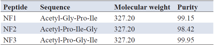
Peptides were synthesized using solid phase organic synthesis technology by Leon (Nanjing) Biotechnology Co. We generated three proline containing tripeptides: NF1: acetyl-Gly-Pro-Ile (acetyl-GPI); NF2: acetyl-Pro-Ile-Gly (acetyl-PIG); and NF3: acetyl-Pro-Gly-Ile (acetyl-PGI). We also synthesized a previously characterized proline-containing dipeptide acetyl-Pro-Ile (acetyl-PI) and palmitoyl pentapeptide-4 to use as positive controls [15, 16]. All peptides, except palmitoyl tripeptide-1, were acetylated at their N-terminus. The purity of the synthesized peptides was assessed by high-performance liquid chromatography (HPLC), with all peptides being more than 98% pure, and their molecular identify was confirmed by mass-spectrometry (Table 1). The fibroblast growth factor receptor (FGFR) inhibitor PD173074 was purchased from Stem Cell Technologies Inc. (Cat #72164). Veliparib (a poly(AD-Pribose) polymerase (PARP) inhibitor) was purchased from MedChemExpress (Cat #HY-10129).

2.2 Cell proliferation assay
Human dermal fibroblast (HSF) and human keratinocyte (HaCaT) cells were obtained from the National Collection of Authenticated Cell Cultures (Shanghai, China), HSF and HaCaT cells were maintained in Dulbecco’s modified Eagle high-glucose media (DMEM, Life Technologies Inc., Cat #C11965500BT), containing 10% fetal bovine serum (FBS), 100 U/ml penicillin, and 100 μg/ml streptomycin. Cells were cultured at 37 °C under 5% CO2 with saturated humidity in an incubator. Cells growing in logarithmic phase (viability >95%) were used in the experiments.
To assess cell proliferation, 6×10⁴ (6,000) cells in a volume of 100 μl were plated into wells of a 96-well plate in the above media and allowed to adhere overnight. The media was then replaced with DMEM with 0.5% FBS. Peptides at final concentrations of 0 (control), 25, 50, and 100 μg/ml were added, with 6 replicates for each treatment and incubated for 48 h at 37 °C. Cell numbers were measured after 48 h using the CCK8 method (Cell Counting Kit-8; MedChemExpress, Cat # HY-K0301), where 10 μl of the CCK8 reagent was added to each well, incubated at 37 °C for 1 h, and the absorbance (OD) measured at 450 nm with a microplate reader.
To assess the role of the FGF receptor pathway in the action of the tripeptides in HSF fibroblast cells, the FGF receptor inhibitor PD173074, at final concentrations of 0, 8.33, 16.27, 33.33, 100, 300, 600, and 1,200 nM, was added to culture media together with the tripeptides NF2 and NF3, at a final concentration of 100 μM, along with the control dipeptide acetyl-PI that had previously been shown to induce skin cell growth through the FGF receptor, and processed as described above except that the cells were grown for 72 h [15]. Cell numbers were measured using the CTG method, where 50 ml of CellTiter-Glo reagent (Promega Inc., Cat #G7570) was added to each well, mixed, and incubated at room temperature for 30 min. Chemiluminescence (RLU) values were measured with an Envision plate reader (Revvity Inc).
To measure the anti-aging effect of the tripeptides, cells were cultured in 12-well plates with 1 ml of cells at a density of 1×10⁵ per ml. After allowing the cells to adhere to the plates, media was replaced with DMEM with 0.5% FBS. Peptides at final concentrations of 0 (control) 10, 20, and 50 μg/ml were added, with 3 replicas of each treatment, and incubated for 24 h. Culture media was then replaced with DMEM with 0.5% FBS and 10 or 20 μM veliparib and incubated for an additional 24 h to induce senescence. Senescent cells were detected using a kit from Beyotime Biotechnology Co. (Cat #C0602). Culture media was aspirated, and cells were washed with PBS and 1 ml of β-gal fixative was added to each well and incubated for 15 min at room temperature. Cells were then washed 3 times with PBS, with 1 ml of staining solution then added and allowed to incubate at 37 °C in the absence of CO2 for 12–24 h. Senescent cells were visualized under an optical microscope.
2.3 Real-time reverse transcriptase quantitative polymerase chain reaction (RT-qPCR)
To measure the influence of our tripeptides on the expression of genes, cells were cultured as above at a density of 2.5×10⁵ cells per well in a 6-well plate, with 2 ml of cells for HSF and 3 ml of cells for HaCaT. After 48 h of culture with tripeptides, cells were lysed by the Trizol method, with chloroform extraction and RNA precipitation with isopropanol. RNA was dissolved in RNAse-free water, and the concentration and purity were assessed using a UV spectrophotometer. 1 μg of RNA was used as template for reverse transcription using a RT Master Mix for qPCR (gDNA digester plus) kit (MedChemExpress, Cat #HY-K0511). Quantitative PCR was conducted using in a 20 μl reaction volume that included 10 μl of 2 X SYBR Green Master Mix (Tiangen Biotech (Beijing) Co., Cat #FP205), 0.3 μM (0.6 μL) each primer (see Table 2 for primers), 2 μl (~50–100 ng) of cDNA and 6.8 μL ddH2O. Reaction conditions included an initial denaturation at 95 °C for 30 seconds followed by 40 cycles of 95 °C for 5 seconds and 60 °C for 30 seconds on a LightCycler® 480 II (Roche). Primer specificity was verified by melting curve analysis and the relative expression level of the transcripts was assessed using the 2-ΔΔCt method, with Glyceraldehyde-3-phosphate dehydrogenase (GAPDH) used as an internal reference [17].
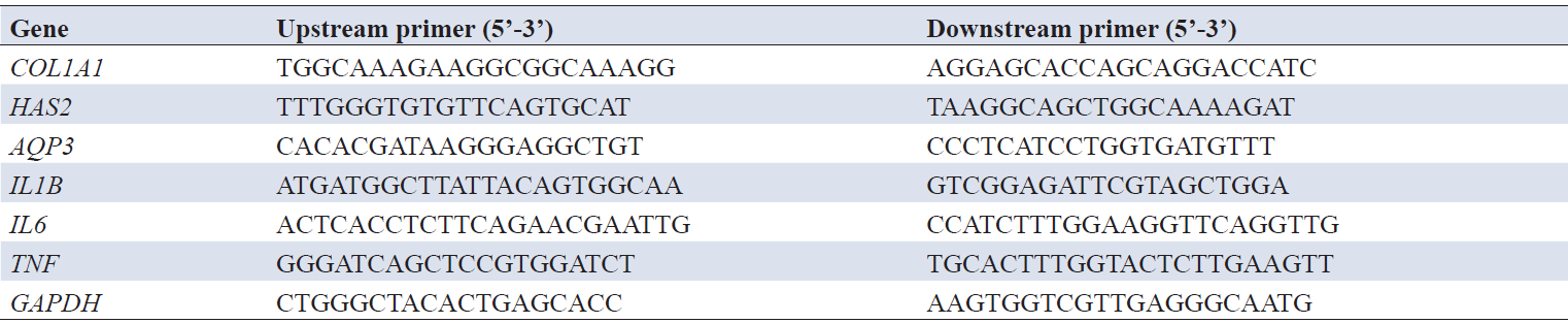
Note: RT-qPCR, reverse transcriptase real-time quantitative PCR; COL1A1, type 1 collagen; HAS2, hyaluronic acid synthase 2; AQP3, aquaporin 3; IL1B, interleukin 1-beta; IL6, interleukin 6; TNF, tumor necrosis factor-alpha; GAPDH, glyceraldehyde-3-phosphate dehydrogenase.
2.4 Epidermis permeability assay
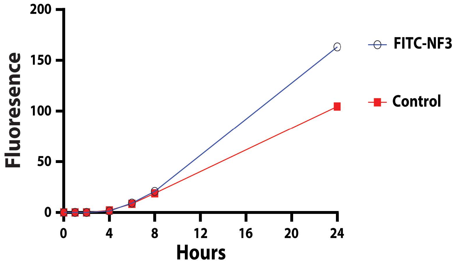
Fluorescein Isothiocyanate (FITC)-labeled NF3 was synthesized by GL Biochem (Shanghai) Ltd. The permeability of FITC-labeled tripeptide was tested using pig skin obtained from Aperture Biotechnology (Shandong) Co. (Cat # TPSY-22). Briefly, labeled tripeptide was applied to the top of skin in a diffusion pate, incubated at 32 °C for 24 h, with samples of the lower media reservoir sampled at 1, 2, 4, 8 and 24 h (with replacement with an equal volume of fresh media). The fluorescence of the sampled media was assessed using a spectrophotometer.
2.5 Statistical analysis
All data are presented as mean ± standard error (SE). Statistical analyses were performed using unpaired t-tests. t-tests were conducted using the “T test calculator” provided at https://www.graphpad.com/quickcalcs/ttest1/?format=c. P values of <0.05 was considered as statistically significant.
3.1 Proline-containing tripeptides promote skin keratinocyte and fibroblast cell growth
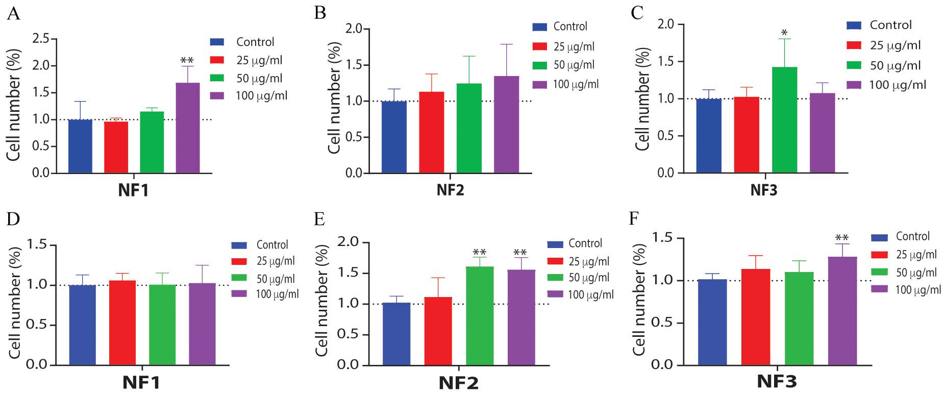
Previous studies have shown that some short peptides stimulate the growth of skin fibroblast and keratinocyte cells [14, 15, 18]. Here we tested 3 proline-containing tripeptides (NF1 (acetyl-GPI), NF2 (acetyl-PIG), and NF3 (acetyl-PGI)) for their effect on the proliferation of HSF cells, a skin fibroblast cell model, and HaCaT cells, a skin keratinocyte cell model [19, 20]. As shown in Figure 1A-C all three proline-containing tripeptides increased the proliferation of HSF fibroblast cells, with NF1 yielding the greatest significant increase in proliferation at the highest concentration (100 μg/ml), NF2 having a concentration-dependent response, and NF3 having a maximal significant response at an intermediate concentration (50 μg/ml). With HaCaT keratinocyte cells, NF1 (Figure 1D) failed to induce a significant increase in proliferation however, tripeptides NF2 (Figure 1E) and NF3 (Figure 1F) significantly increase proliferation, with NF2 having the largest increase. The effects of the three tripeptides on the two cell lines were unique, with NF1 having the largest increase, seen in HSF cells, of any tripeptide in either cell line, yet resulting in no increase in proliferation of HaCaT cells. NF2 caused a larger increase in HaCaT cells than in HSF cells (compare Figure 1B, 1E). NF3 increased proliferation only at an intermediate concentration (50 μg) in HSF cells (Figure 1C) and had a moderate effect on HaCaT cells (Figure 1F). These results suggest that the proline-containing tripeptides NF1, NF2, and NF3 increase the proliferation of skin cells, both fibroblast and keratinocytes, but that they have variable and unique effects.
3.2 FGF receptor activity inhibitors block the growth prompting action of the proline-containing tripeptides on skin fibroblast cells
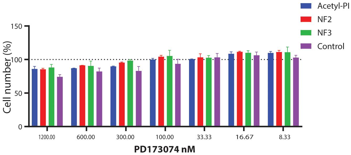
Skin fibroblast cell proliferation can be regulated through the FGF receptor signaling pathway, as has been shown for some short peptides, e.g., dipeptide acetyl-Pro-Ile [11, 15]. To determine whether our tripeptides also induce proliferation through the FGF receptor pathway we tested two of our tripeptides (NF2 and NF3) with increasing doses of a FGF receptor pathway inhibitor, PD173074. As shown in Figure 2, at low concentrations of the PD173074 FGF receptor inhibitor (8.33 and 16.67 nM) tripeptides NF2 and NF3, along with the positive control dipeptide acetyl-Pro-Ile increase proliferation of HSF fibroblast cells, but at higher concentrations of the inhibitor PD173074 the increase in proliferation rates is abolished. These results suggest that the tripeptides NF2 and NF3 at least partially act through the FGF-receptor signaling pathway.
3.3 Proline-containing tripeptides upregulate the abundance of type I collagen (COL1A1) mRNA transcripts in skin cells
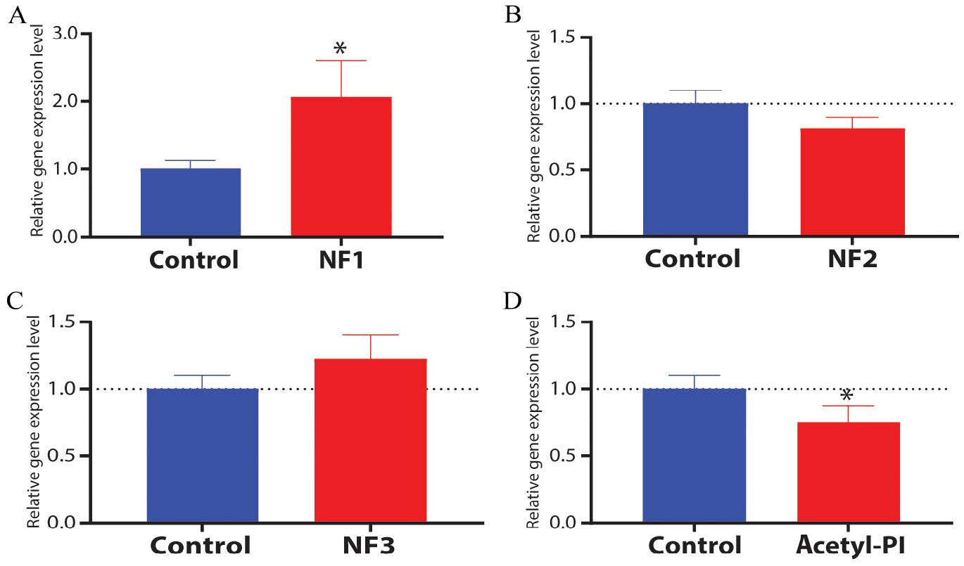
Collagen is an important protein component of skin, providing strength and elasticity [21]. To determine whether our tripeptides increase the abundance of the type I collagen (COL1A1) mRNA we used reverse transcriptase real-time quantitative PCR (RT-qPCR) in HSF skin fibroblast cells (Figure 3). As shown in Figure 3A tripeptide NF1 significantly increased COL1A1 mRNA abundance, with mRNA abundance approximately doubling. Tripeptide NF3 yielding about a 20% increase (Figure 3C), which was not significant, while tripeptide NF2 resulted in no increase (Figure 3B). The dipeptide acetyl-Pro-Ile did not increase COL1A1 mRNA abundance, instead, produced to a significant decrease in mRNA abundance (Figure 3D). The increase in COL1A1 mRNA abundance due to tripeptide NF1 was greater than this tripeptide’s impact on cell proliferation (Figure 1A), thus we conclude that there was an increase in COL1A1 mRNA number per cell. For tripeptide NF3, the increase in COL1A1 mRNA abundance was similar to fold change in cell proliferation (Figure 1C), thus the change in COL1A1 mRNA abundance may simply be due to an increase in cell number. From this experiment we concluded that exposure to tripeptide NF1 increases COL1A1 mRNA abundance in HSF skin fibroblast cells.
3.4 Proline-containing tripeptides upregulate the abundance of moisture-related and inflammation-related gene mRNAs in skin fibroblast cells
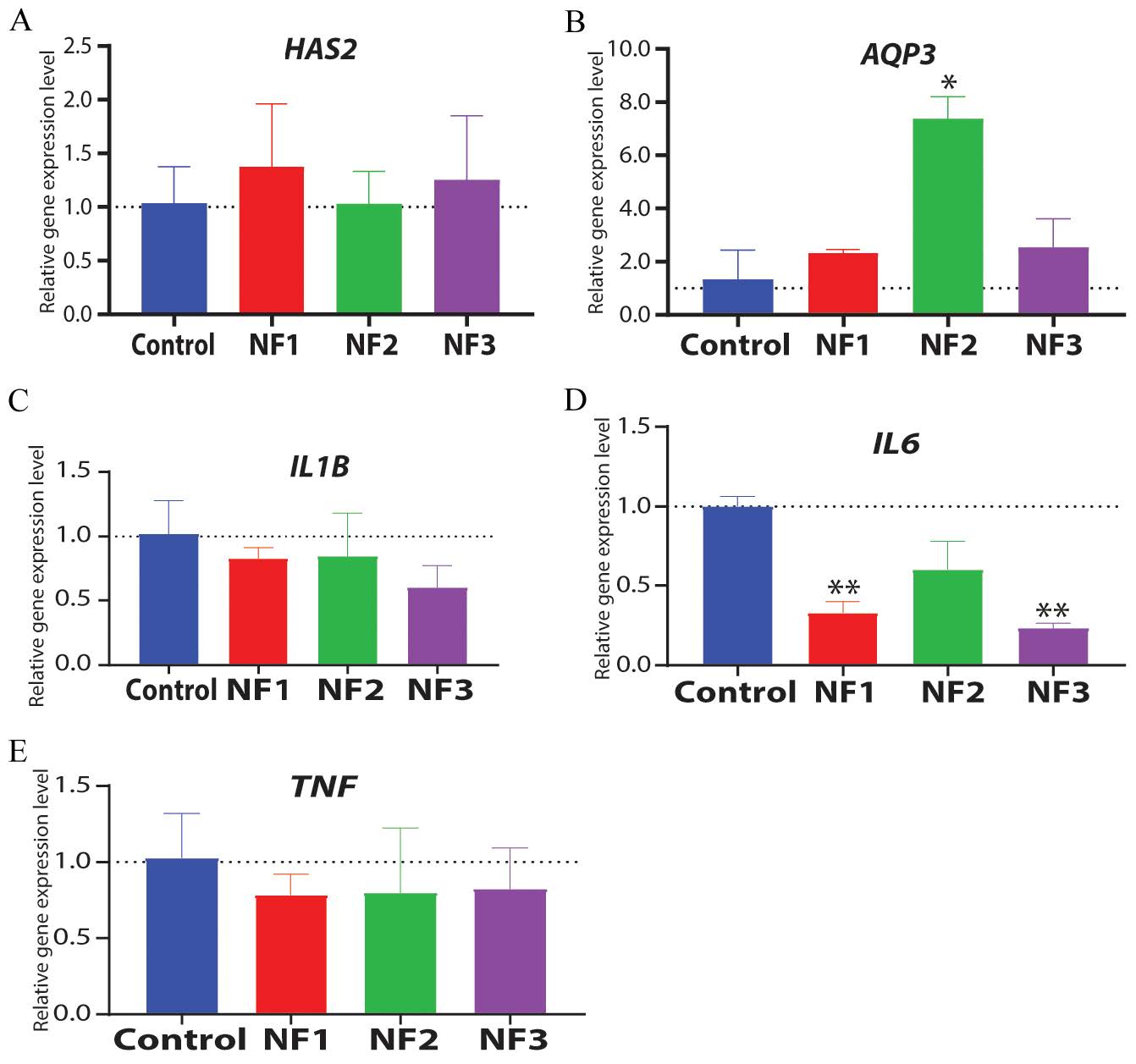
Changes in the mRNA abundance of additional genes in the skin cells influence skin properties such as moisture content (hydration) and inflammation, with fibroblasts playing a key role in these processes [22, 23]. To examine this, we used reverse transcriptase real-time quantitative PCR (RT-qPCR) to quantify the transcript abundance of 2 genes associated with moisture content of skin, HAS2 (hyaluronic acid synthetase 2) and AQP3 (aquaporin 3), and 3 genes associated with inflammation, IL1B (interleukin 1 beta), IL6 (interleukin 6), and TNF (tumor necrosis factor–alpha) in HSF skin fibroblast cells. As shown in Figure 4, tripeptides NF1 and NF3 induce a modest, but not significant, increase in the abundance of the HAS2 transcript (Figure 4A). Tripeptide NF2 generated an approximately 7-fold significant increase in AQP3 transcript abundance, with more modest, and not significant, increases seen with tripeptides NF1 and NF3 (Figure 4B). The significant 7-fold increase in AQP3 transcript abundance with tripeptide NF2 far exceeded the increase in cell numbers. The tripeptides generally reduced the abundance of inflammation-related gene transcripts, with greatest reductions (significant with P<0.01) seen for IL6 when exposed to tripeptides NF1 and NF3 (Figure 4C-E). Modest, non-significant, down regulation of IL1B mRNA abundance, with tripeptide NF3 having the strongest effect (Figure 4C), and TNF mRNA (Figure 4E), with all tripeptides having a similar effect, was observed. For IL6, tripeptide NF3 had the strongest effect, with tripeptide NF2 leading the smallest down regulation (both significant, P<0.01; Figure 4D). These results show that each proline-containing tripeptide has a unique effect on the expression of genes associated with moisture content and inflammation.
3.5 Proline-containing tripeptides reverse the effect of aging on gene expression in skin cells
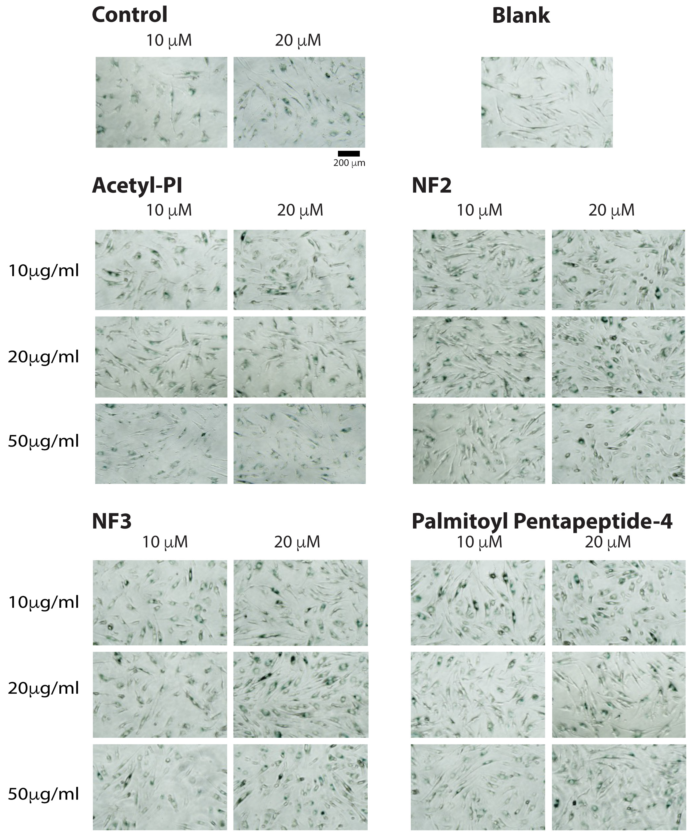
Skin ages, and the environment can induce premature aging [2]. We tested whether our proline-containing tripeptides could reduce or reverse the aging phenotype of fibroblast cells. HSF fibroblast cells were pretreated for 24 h with our tripeptides prior to treatment with the aging agent Veliparib, which mimics the effects of aging, for an additional 24 h [24]. As shown in Figure 5, pretreatment with all tripeptides dose-dependently reduced the number of senescent cells, when cells were treated with Veliparib at both 10 μM and 20 μM. These results suggest that our proline-containing tripeptides can protect skin fibroblast cells from the aging effects of Veliparib.
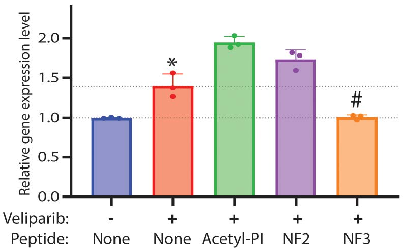
To better understand how the tripeptides protect the skin cells from aging, we examined MMP1 (matrix metalloprotease 1) gene expression (Figure 6). Veliparib induces a significant increase in MMP1 mRNA abundance. Tripeptide NF3 was found to significantly restore basal levels of MMP1 gene expression, reversing the effects of Veliparib, in contrast to tripeptide NF2, which did not reduce MMP1 mRNA abundance. These results indicate that the proline-containing NF3 may prevent the harmful effects of Veliparib on HSF fibroblast cells.
3.6 A proline-containing tripeptide can penetrate the epidermis in a pig skin model
Since skin acts as a barrier to prevent many compounds from entering an individual, we sought to determine whether one of our short proline-containing tripeptides could penetrate skin [1]. Here we tested the ability of FITC-labelled NF3 to penetrate skin using a pig skin model. As shown in Figure 7, increased amounts of FITC-label could be detected in the lower receiver media of skin treated with FITC-labeled NF3 after incubation for 8 or more hours. Even after 24 hours the pig skin retained its integrity. This result suggests that short proline-containing tripeptides potentially can penetrate skin and thus may act on keratinocytes and fibroblasts within the epidermis and dermis.
Fibroblast growth factors (FGFs) are important regulators of cell growth in multiple human tissues, including skin, and have been considered as a therapy to treat skin tissue damage [9, 25-28]. Several FGF proteins are known to be expressed in skin tissue to modulate cell growth and tissue repair [9, 26]. FGF7 is used as a therapy to treat skin burns and wounds, but this is only possible as this large protein has access to the damaged tissue [28]. For other types of skin damage, such as aging, therapies with intact FGF proteins are not possible as the cells that need to be targeted as protected by other skin cells, which prevent large molecules from penetrating into the tissue [29]. Great efforts have been made to identify small molecules, and improved methods that allow molecules to penetrate skin, to deliver therapeutic or cosmetic benefits [29, 30]. A potential solution to this problem is to identify short peptides that mimic the action of larger molecules. This has been accomplished by testing a diversity of peptide starting materials [e.g., 14] or by more directed studies designing peptides derived from these larger proteins [12].
FGF proteins exert their effect on cells by binding to fibroblast growth factor receptors (FGFRs) [9]. Studies have identified amino acid residues in both FGFs and FGFRs that are responsible for ligand-receptor binding [9, 10]. With the identification of these amino acid residues several groups have synthesized peptides that correspond to these sites, such as canofins, which are 8–11 amino acids long, and the dekafins, which are 14 amino acids long, to determine whether these peptides can induce receptor signaling [11-13]. While some of these peptides function to mimic FGF activity they are still too long (8–14 amino acids) to penetrate skin [12, 13]. Alternative approaches identified a short peptide (GPIGS), which was derived from a bacterial source that promotes keratinocyte growth [14]. Subsequent studies showed that a short dipeptide, Ile-Pro, derived from this sequence retains keratinocyte growth-promoting activity that mimics FGF and is skin permeable [15]. Comparison of this dipeptide sequence to known interaction sites between FGFs and FGFRs suggests that a conserved proline residue in the FGF sequences adjacent to the β12 loop could be part of an FGF-FGFR binding site [13]. This observation suggested to us that the β12 loop might be a promising therapeutic candidate for developing skin permeable proline-containing tripeptides to modulate FGFR activity in skin tissue.
Here, we characterize several proline-containing tripeptides and show that all three of our tripeptides induce proliferation of skin cell models, with NF1 and NF3 promoting HSF skin fibroblast cell growth and NF2 and NF3 promoting cellular proliferation of the HaCaT skin keratocytes (Figure 1). These results are in accord with those reported for other larger peptides derived from FGF sequences [12, 13]. When we used an inhibitor of FGF signaling (PD173074) we impaired the ability of skin HSF fibroblast cells to increase their growth rates in response to our proline-containing tripeptides (Figure 2). However, we did not show, e.g., by Western blot of downstream effectors, that our peptides impair FGFR signaling. This result suggests that our proline-containing tripeptides act, at least partially, the through the FGF signaling pathway, potentially by mimicking the β12 loop of FGF to bind and activate FGFRs. Further studies are needed to confirm the identify of specific FGFRs involved in this process, and the specific sites within the receptors mediating this effect.
While cell growth is important, we sought to determine whether our proline-containing tripeptides also influence other phenotypes of skin cells that might be beneficial for skin tissue. Collagen, which is synthesized by skin fibroblast cells, is an essential component of the extracellular matrix of skin tissue and is necessary for skin tissue function [31]. We found that only one of our three proline-containing tripeptides, NF1, induced a significant increase in COL1A1 mRNA abundance, and likely collagen protein production, in skin HSF fibroblast cells (Figure 3). The increase in COL1A1 mRNA abundance with tripeptide NF1 (Figure 3) was much larger than the increased cell proliferation (Figure 1), thus we conclude that NF1 caused an upregulation of COL1A1 mRNA in skin HSF fibroblast cells. In contrast, the dipeptide PI, which previously was shown to induce keratinocyte growth and act through FGFRs significantly decreased COL1A1 mRNA abundance (Figure 3), a property that is not beneficial for skin repair [15].
Healthy skin contains water that increases the volume of skin tissue and reduces the appearance of wrinkles [32]. As skin ages or is injured, it loses its ability to hold water and wrinkles develop [33]. When fibroblast cells were treated with our proline containing tripeptides, only modest increases in HAS2 mRNA abundance were observed, however a significant seven-fold increase in AQP3 mRNA abundance was seen with the tripeptide NF2 (Figure 4A, 4B). Previous work has shown that AQP3 gene expression is regulated by FGF2 in breast cancer, thus our results are not unprecedented and suggest that the regulation of aquaporin genes by FGFs may be more widespread [34]. AQP3 encodes a water transporter, thus increased AQP3 mRNA levels should increase AQP3 transporter levels and thus increase water uptake by fibroblast cells [35]. Increased water uptake by fibroblast cells should increase their volume, along with skin tissue volume, and potentially reduce skin wrinkling.
An important function of skin is to act as a barrier to prevent infection [32, 36]. As skin ages, this barrier function weakens, allowing the entry of molecules that induce immune responses such as inflammation [37]. Inflamed skin is not healthy skin. Much of the inflammation is due to immune cell responses to factors, such as interleukins, secreted by skin cells [38]. When skin HSF fibroblast cells were treated with our three proline-containing tripeptides, modest reduction in the abundance of IL1B and TNF mRNA transcripts were seen with all three tripeptides, and more importantly a large significant reduction in IL6 transcript levels was observed (Figure 4C-E). This result parallels the observation that FGF-2 can decrease the production of interleukin-6 [37]. The downregulation of the three immune related transcripts that we examined should lead to reduced production of these pro-inflammatory cytokines and thus reduced inflammation of skin, and improved appearance.
Therapies that treat skin conditions are useful, but preventative agents might be better for skin health [36, 37]. Matrix metalloprotease abundance increases with skin damaging and aging, thus can be used as a biomarker [39]. Our experiments show that pre-treating skin HSF fibroblast cells with our proline-containing tripeptides reduce senescence of cells when treated with an aging compound Veliparib (Figure 5), and at least one of them, NF3, significantly reduces the abundance of MMP1 mRNA, a biomarker of aging, in these cells (Figure 6).
A limitation of many potential therapeutic agents for improving skin health is that they cannot be effectively delivered to skin cells due to their size or hydrophobicity [29, 30]. Small and hydrophobic molecules have greater potential to penetrate skin [29, 30]. Previous work has shown that some short peptides, with lipid modifications can penetrate skin, and thus target cells within the epidermis and dermis [15]. We assessed the potential of one of our tripeptides to penetrate skin using a transdermal assay. As shown in Figure 7, one of our tripeptides was seen to pass through skin and accumulate on the other side of a transdermal assay. The greatest increase is seen at 24 h, with a small increase at 8 h, indicating that stable molecules need to be used for this type of therapy. We suspect that the accumulation of the FITC label is due to passive diffusion as we have no evidence for active transport. However, further studies are needed to confirm the generality of the potential of short proline-containing peptides to penetrate skin, and the mechanism by which they do so.
In conclusion, this work shows that some of our proline-containing tripeptides possess properties that are beneficial for skin health. They promote the growth of both keratinocyte and fibroblast cells, properties that have been found in other FGF mimetics, and that this action is mediated at least in part through FGFRs. The tripeptides also increase the abundance of extracellular matrix and moisture related gene transcripts, and suppress transcripts for pro-inflammatory molecules, processes that are also regulated by FGFs. The tripeptides also suppress senescence, and act as preventative agents to block damage. Importantly, unlike longer FGF mimetics, we showed that one of our short tripeptides can penetrate skin, thus target the cells required to improve skin biology. No single proline-containing tripeptide possessed all of the desirable properties, but an effective therapy to improve skin health may be developed using a mixture of these tripeptides.
AQP3, Aquaporin 3; COL1A1, Type 1 collagen; FGF, Fibroblast growth factor; FGFR, Fibroblast growth factor receptor; FITC, Fluorescein Isothiocyanate; GAPDH, Glyceraldehyde-3-phosphate dehydrogenase; HAS2, Hyaluronic acid synthase 2; IL1B, Interleukin 1-beta; IL6, Interleukin 6; MMP1, Matrix metalloprotease 1; RT-qPCR, Reverse transcriptase real-time quantitative PCR; TNF, Tumor necrosis factor-alpha; HSF, Human skin fibroblast; PI, Proline-isoleucine (dipeptide).
Author contributions
ZT, MWI, and QZ contributed to the conception of the study and contributed significantly to analysis. DMI contributed to the conception of the study, contributed significantly to analysis and manuscript preparation, and wrote the manuscript.
Funding
This research received no external funding.
Data availability
Data used in this manuscript can be obtained from the authors.
Ethics approval and consent to participate
Not applicable.
Consent for publication
All authors provide consent to publish this manuscript.
Competing interests
MWI and DMI own shares of Shanghai Niefei Biotechnology Co. Ltd.
Acknowledgements
This work was supported by Shanghai NieFei Biotechnology Co. Ltd. The authors wish to thank Ken Xing for technical assistance.
ISSN: 3105-7713
Volume 1, Issue 1
15 June – 14 September
Pages: 1 – 65


 Log In

 On This Page
 CITE
CITE