 Log In
Search




 Open Access
 zengqingyu2011@tongji.edu.cn
Qingyu Zeng, Institute of Photomedicine, Shanghai Skin Disease Hospital, School of Medicine, Tongji University, No. 1278 Baode Road, Jing'an District, Shanghai 200443, China. E-mail: zengqingyu2011@tongji.edu.cn.
Open Accesszengqingyu2011@tongji.edu.cnQingyu Zeng, Institute of Photomedicine, Shanghai Skin Disease Hospital, School of Medicine, Tongji University, No. 1278 Baode Road, Jing'an District, Shanghai 200443, China. E-mail: zengqingyu2011@tongji.edu.cn.
Spatial transcriptomics (ST) preserves tissue structure and maps gene expression in physical space. Unlike bulk RNA-seq or scRNA-seq, ST keeps positional information [1, 2]. Because skin function depends directly on its layered architecture, it is a tissue for which dissociative transcriptomic approaches are inherently limited. The clinical and biological heterogeneity of skin diseases therefore makes the case for applying ST particularly strong.
ST captures transcriptomic information at the spot level or even at near-single-cell resolution directly in situ and links each measurement to a spatial coordinate, enabling the simultaneous interrogation of gene expression profiles and tissue spatial architecture. Current ST platforms fall into two categories: imaging-based and sequencing-based methods [3]. The skin contains diverse cell types and varies markedly across patients. ST connects morphology with molecular profiles. It can pinpoint the actual locations where disease-related interactions occur. This could offer a practical roadmap for targeted interventions [4].
Skin diseases are heterogeneous across multiple levels. In psoriasis and atopic dermatitis (AD), clinical manifestations and histopathological features differ markedly among patients. At the tissue and cellular levels, the epidermis, dermis, and skin appendages, such as hair follicles and sebaceous glands, are intricately stratified and comprise diverse cell types, including keratinocytes, fibroblasts, endothelial cells, and a wide range of innate and adaptive immune cells [5]. Conventional “homogenization” approaches struggle to answer where inflammatory signals originate, which cells drive them, and how they are amplified and sustained within a given spatial architecture. The application of ST is therefore transforming our understanding of skin disease from a two-dimensional “cellular parts list” into a three-dimensional atlas of the spatial organization of cells [6].
This review focuses on using ST to investigate the pathogenesis and potential therapeutic targets of skin diseases. We first summarize mainstream ST technology routes and key considerations for platform selection. We then outline recent progress enabled by ST technologies in inflammatory skin diseases, autoimmune and fibrotic disorders, and skin tumors, highlighting newly defined spatial niches, cell–cell communication programs, and molecular subtypes. We also discuss how these insights can facilitate the identification of novel targets, the optimization of therapeutic regimens, and the development of strategies to overcome treatment resistance by remodeling pathogenic microenvironments.
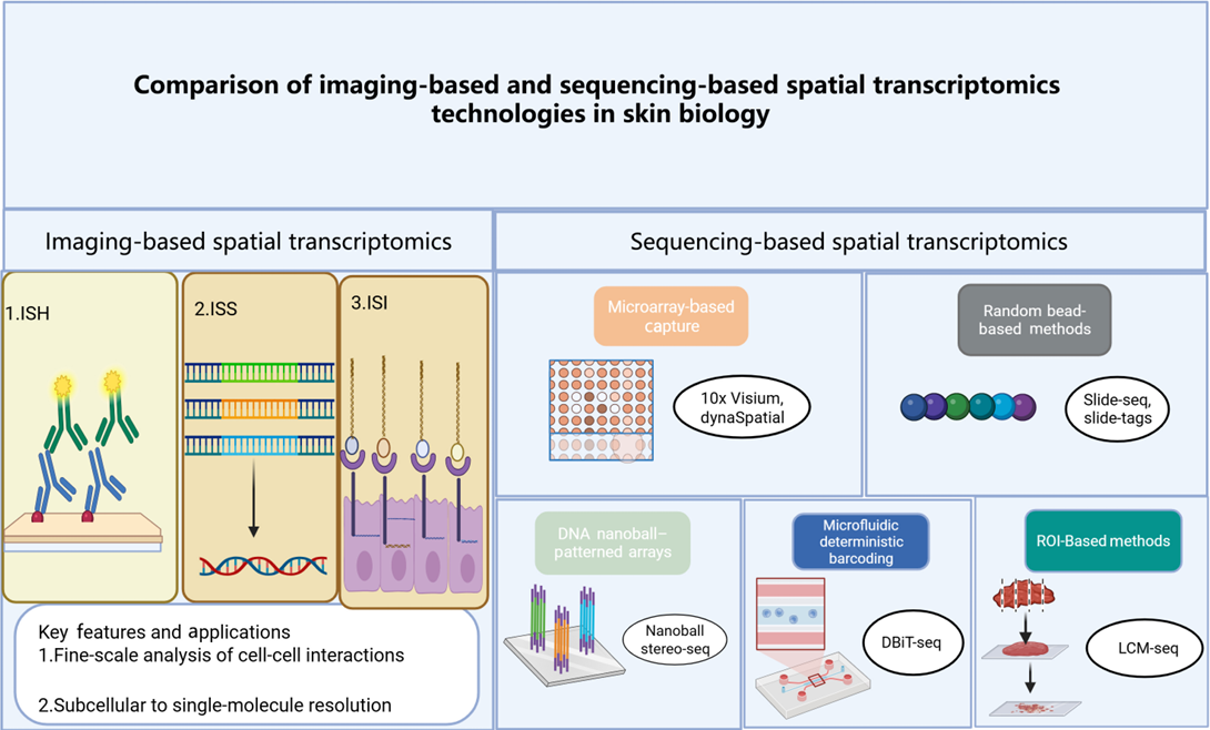
Based on their technical principles and implementation strategies, existing ST methods can be broadly categorized into imaging-based and sequencing-based approaches, as shown in Figure 1. Each category encompasses several techniques with distinct strengths and limitations, which must be care-fully considered for their optimal application in skin disease research.

2.1 Imaging-based ST
Imaging‑based ST methods, such as in situ hybridization (ISH), in situ sequencing (ISS) and in situ imaging (ISI), can detect RNA molecules directly in fixed tissue sections [7-9]. They achieve nanoscale or subcellular resolution and work well with formalin-fixed, paraffin-embedded samples [10]. They can even analyze fine structures such as hair follicle niches [11]. Thus, these methods are particularly well suited for analyzing complex anatomical regions. In skin research, high-resolution spatial data can show gene activity clearly in all skin layers. They can reveal clusters of immune cells near skin glands and hair follicles. They can also detect changes in skin cells. However, imaging‑based methods can detect only a limited number of transcripts and are relatively costly. These limitations restrict the broader application of imaging‑based ST [12].
2.2 Sequencing-based ST
High-definition array-based platforms such as 10x Visium have become widely used tools for spatial profiling [13]. Although Visium HD can achieve ~2 µm resolution, it demands considerable computational resources [2, 14, 15]. Standard 55–100 µm spots reduce the heavy computational burden but do not provide single-cell resolution [13, 16].
Bead‑based methods provide an alternative. Slide-seq and Slide-tags use densely packed barcoded beads spaced 10–20 µm apart to achieve single-cell resolution. These methods can better resolve microenvironments and limit RNA diffusion [17-19].
Deterministic barcoding in tissue for spatial omics sequencing (DBiT‑seq) and similar microfluidic deterministic barcoding methods offer another approach. Rather than prioritizing the highest spatial resolution, they enable the simultaneous analysis of RNA and protein expression [20, 21]. Because skin diseases often involve both the epidermis and dermis, this multimodal analysis can help identify intercellular interactions [22, 23].
Region-of-interest‑based platforms such as NanoString GeoMx DSP represent another strategy. They focus on predefined regions, such as hair follicles or psoriatic plaques, rather than enabling continuous tissue-wide profiling. This targeted design is unsuitable for unbiased discovery [24].
However, selecting an appropriate platform is challenging because each technology has distinct strengths and limitations that can affect data interpretation. For example, although Visium HD provides the highest resolution among array-based methods, the resulting data volume can exceed the capacity of standard computational pipelines. More importantly, the 55-µm spots of conventional Visium arrays mix transcripts from multiple cells. Bead-based platforms partially address this limitation through finer spatial sampling. However, random bead placement does not guarantee true single-cell capture [24].
The choice of a suitable platform depends largely on the disease under investigation. In melanoma, array-based methods may be appropriate because the lesions often have clearly defined tissue boundaries. In psoriasis and acne, where pathological margins may be poorly defined, bead-based approaches can minimize signal crosstalk. DBiT-seq is suitable when simultaneous analysis of RNA and protein expression is required. For well-defined structures such as hair follicles, region-of-interest-based platforms may be appropriate. No single platform is suitable for all research settings. The best choice depends on the specific research question and tissue complexity [12].
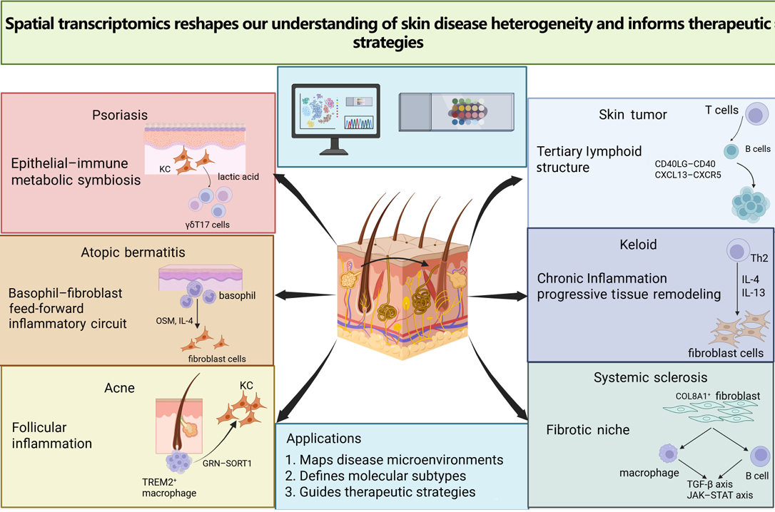
An overview of key spatially resolved mechanisms across major skin diseases is presented in Figure 2.
3.1 Inflammatory skin diseases
3.1.1 Psoriasis
Recent studies using ST have revealed complex cellular circuits that drive psoriasis. This approach has revealed an IL-36 inflammatory hub in the upper epidermis. Specific fibroblast subsets located in the papillary dermis are associated with lesion recurrence [25, 26]. Activated keratinocytes participate in the IL-36 loop to amplify local inflammation [25]. Basal keratinocytes exhibit a metabolically dominant state characterized by COX7B‑driven oxidative phosphorylation combined with glycolytic lactate production. This metabolic state fuels their hyperproliferation and also promotes the expansion of neighboring γδT17 cells. This creates a pro‑inflammatory feedback loop between epithelial and immune cells [27, 28]. A discrete AKR1B10⁺/MT1G⁺ spinous-layer keratinocyte subset has also been linked to disease severity and treatment resistance [29, 30]. Even clinically uninvolved skin in severe psoriasis patients exhibits molecular abnormalities. Keratinocytes in those regions appear pre-activated, with upregulation of FGFR3 and DGAT2. This might point to an underlying defect in barrier integrity [26]. Dermal fibroblasts are also actively involved in the inflammatory process. SFRP2⁺ fibroblasts recruit monocytes and activated dendritic cells. CD8⁺ T cells secrete CCL13, CCL19, and CXCL12 [25]. WNT5A⁺/IL-24⁺ fibroblasts promote matrix remodeling and angiogenesis [31, 32]. FAP⁺ fibroblasts cluster with endothelial cells and antigen‑presenting cells to form structures resembling tertiary lymphoid microdomains [33, 34].
Patients who respond poorly to IL-23 blockade exhibit persistent Th17 activation programs [35]. In contrast, a response to IL-17A blockade with agents such as secukinumab is associated with the downregulation of hypoxia-related pathways involving genes such as RHCG [36]. Although treatments such as secukinumab and Dead Sea climatotherapy can achieve similar clinical effects, they leave distinct molecular scars. These molecular imprints differ in their transcriptomic profiles, with 479 genes, including SERPINB4, IL36G, and AKR1B10, being differentially expressed between the treatment groups. The treatments also produce distinct proteomic changes in key microenvironments, such as CD103⁺ cell niches and the dermis. Such treatment-specific molecular scars may influence disease memory and relapse patterns [37]. Moreover, obesity and resistance to biologic therapy are associated with persistently high expression of epidermal innate immune signals, such as S100A8/A9, IL-36, and TNFSF10/TRAIL. This pattern suggests an innate immunity- and metabolism-driven subtype that is independent of classical Th17 pathways [38]. Together, these findings from ST define a spatially organized inflammatory circuit. This circuit spans epidermal amplifiers, metabolically primed keratinocytes, and heterogeneous fibroblast hubs.
3.1.2 AD
The combination of ST and single-cell RNA sequencing has revealed cellular circuits involved in AD [39]. These techniques localize disease-associated cellular interactions to specific regions of the skin, thereby helping to guide therapies targeting specific tissue regions [40-42].
Keratinocyte hyperproliferation contributes directly to two key features of AD: epidermal thickening (acanthosis) and dysregulated keratinization. ST enables the localization of disease-associated changes to specific tissue regions. Activated keratinocytes produce large amounts of chemokines such as CCL17 and CCL22 and cytokines like TSLP and IL-33. These mediators recruit and activate immune cells, thereby sustaining inflammation in AD [43]. Barrier dysfunction is shown at the molecular level. At the same time, IL-4 and IL-13 derived from Th2 cells directly downregulate key barrier-related genes, such as FLG, which encodes filaggrin. Additionally, IL-22 secreted by Th22 cells acts synergistically with Th2 cytokines to induce spongiosis and differentiation defects characteristic of AD [44, 45].
In the dermis, fibroblasts serve as key amplifiers and form active signaling networks with multiple immune cell populations. In leukocyte-infiltrated regions, COL18A1-expressing activated fibroblasts co-localize with CCR7-expressing dendritic cells, CCL13/CCL18-expressing M2 macrophages, and T cells, establishing stable communication networks via ligand–receptor pairs such as CCL19–CCR7 [42]. Deeper mechanisms have been revealed in mouse models. Basophils can initiate a potent basophil–fibroblast feed-forward inflammatory circuit by secreting oncostatin M and IL-4. This circuit provides potential targets for disrupting pathogenic intercellular communication [43, 46].
ST has revealed extensive remodeling of immune cell compartments in AD lesions. Macrophages and dendritic cells increase markedly in abundance and serve as bridges between innate and adaptive immunity. Their infiltration is a hallmark of immune activation, and these cells serve as major sources of inflammatory mediators, secreting cytokines such as TNF-α and IL-23 that amplify inflammation and may promote distinct inflammatory endotypes, such as Th17- and Th22-associated endotypes. As terminal effectors, Th2, Th22, and Th1 subsets produce IL-4, IL-13, IL-22, and IFN-γ to directly mediate itch, barrier disruption, and epidermal hyperplasia—consistent with AD being fundamentally a T-cell-mediated immune disease [43]. Sebaceous glands are also being redefined as active regulators in disease. ST indicates that sebaceous glands in AD lesions undergo Th2 inflammation-associated lipid metabolic reprogramming, characterized by altered expression of genes such as CCL17 and HSD3B1, suggesting that they may participate in disease-specific local immune modulation [47].
In-depth analysis of multi-omic data has identified new candidate regulators and endotype markers. At least three core AD subtypes have been resolved: (i) a barrier-defect/metabolic-dysregulation subtype characterized by downregulation of UGT3A2 and impaired barrier repair; (ii) an immune-imbalance “endogenous” subtype associated with high CHRM4 expression and low IgE levels, suggesting atypical Th2 activation; and (iii) a cytokine-driven inflammatory activation subtype featuring high expression of IL-36A, IL-4R, and other key factors that drive strong local inflammation. These subtypes point to distinct targets for precision intervention—barrier repair, innate immune modulation, and targeted anti-cytokine therapy—providing a scientific basis for moving from a single clinical diagnosis toward stratified therapy guided by molecular subtyping [43]. These insights have profound implications for treatment strategy. The limitations of strategies that target only the Th2 axis, such as treatment with anti-IL-4Rα antibodies, may be explained by their limited effects on parallel pathways, such as the IL-22 pathway [45]. Therefore, multipathway strategies, such as dual targeting of the AhR and JAK pathways or interventions targeting specific intercellular circuits such as the basophil–fibroblast axis, appear more promising [41, 43, 45, 46]. Collectively, these spatial and single-cell insights are transforming our understanding of the complexity of AD. They reveal distinct disease subtypes and multicellular interaction networks that pave the way for subtype-specific, multitarget precision therapies.
3.1.3 Acne
Sebaceous glands are dynamic and heterogeneous functional units. ST is updating our understanding of their role in acne. By combining single-cell sequencing with ST, studies have distinguished keratinocyte subpopulations across different anatomical regions at the molecular and spatial levels. These studies have also mapped the full differentiation trajectory of human sebaceous lineage cells from proliferation and lipid synthesis to terminal rupture, and defined stage-specific marker genes [48, 49]. These findings enable the precise localization of disease-associated changes.
In acne, immune cells gather at specific microanatomical sites, forming organized clusters that amplify local inflammation. ST reveals that early lesions feature TREM2+ macrophages activated by lipid signals. These cells form a key hub for the initiation of inflammation [50]. Spatial co-localization further suggests direct macrophage–keratinocyte communication through axes such as GRN–SORT1, promoting keratinocyte hyperproliferation and hyperkeratinization and thereby providing a spatial explanation for the cellular origin of follicular hyperkeratosis and comedone formation [51, 52]. ST identifies a scar-associated niche in which profibrotic SPP1+ macrophages coexist with POSTN+ fibroblasts and cooperate through SPP1 signaling to drive abnormal collagen deposition [53].
Based on these mechanistic insights, TREM2+ macrophages and downstream molecules such as SORT1 and SPP1 have emerged as potential therapeutic targets to interrupt acne inflammation and fibrotic progression [50, 53]. Equally important, ST provides a powerful framework to evaluate how existing therapies remodel the spatial ecology of lesions.
3.2 Skin tumors
3.2.1 Cutaneous squamous cell carcinoma (cSCC)
The pathogenesis of cSCC is driven by genetic mutations, tumor heterogeneity, and remodeling of the immune microenvironment. Spatial omics reveals that driver mutations in TP53 and NOTCH1 initiate the transition from precancerous states to invasive carcinoma, challenging conventional linear progression models [54]. Tumor heterogeneity in skin cancers is largely attributable to keratinocyte plasticity. Keratinocyte differentiation programs depend on signals from the surrounding tissue [6, 55, 56]. Cancer-associated fibroblasts (CAFs), for instance, promote tumor invasiveness via the MDK–ITGA6 signaling axis. Certain tumor clones also recruit regulatory T cells through CXCL16/CXCR6 chemokine signaling to build an immunosuppressive microenvironment [57, 58]. To target the tumor microenvironment in cSCC, researchers recently developed a modified chlorin e6 derivative called Shengtaibufen. Shengtaibufen induces STING-mediated PANoptosis, when combined with lymphocyte-activation gene 3 blockade, enables a synergistic photodynamic therapy–immunotherapy strategy that reshapes the tumor immune microenvironment [59]. Together, these findings link cSCC progression to spatially coordinated interactions among genetic mutations, stromal cells, and immune populations.
3.2.2 Melanoma
Melanoma heterogeneity is not randomly distributed. An early ST study of lymph node melanoma metastases revealed that distinct tumor subclones occupy separate spatial domains within a single lesion, with MITF-low and MITF-high transcriptional programs intermixed [60].
For example, in melanomas arising from congenital melanocytic nevi, deeper tumor invasion is associated with higher expression of immunosuppressive genes, stronger T-cell inhibitory signatures, and greater M2 macrophage polarization. Metastatic lesions also upregulate additional immune evasion genes like AXL, EPHA2, and BST2. A stress-like cancer cell state characterized by FOS/JUN pathway activation and heat-shock protein expression has also been confirmed in both zebrafish and human melanoma models. This state, which has been observed in multiple cancer types, may contribute to drug resistance [61].
Integrative lineage tracing and ST have established a hierarchical melanoma growth model with tumorigenic competence confined to a perivascular niche and mesenchymal-like cells identified as metastasis-initiating cells [62]. In melanoma brain metastases, multi-omic analyses revealed chromosomal instability, a neuronal-like state, and enrichment of dysfunctional TOX⁺ CD8⁺ T cells and monocyte-derived macrophages [63].
A conserved “interface” cell state at tumor boundaries was identified in zebrafish models, marked by coordinated cilia gene upregulation [64]. In acral melanoma, MYC⁺ melanoma cells and FGFBP2⁺ natural killer T cells were linked to lymph node metastasis through MITF-induced fatty acid oxidation, a targetable dependency [65]. Together, these studies highlight the power of ST to resolve melanoma's cellular heterogeneity, immune remodeling, and therapeutic vulnerabilities.
3.2.3 Basal cell carcinoma (BCC)
Recent advances in single-cell RNA sequencing and ST have provided high-resolution insights into the cellular heterogeneity and spatial organization of skin cancers, particularly BCC [66]. Integrated analyses of human BCC have revealed that tumor cells within the invasive niche adopt collective migration phenotypes characterized by increased expression of cell–cell junction-related genes, whereas spatially adjacent CAFs exhibit extracellular matrix-remodeling features and respond to tumor-derived activin A signaling [67]. A distinct CAF subset (C01_TNC) enriched in peritumoral regions has been identified. It may promote tumor progression through matrix remodeling and spatial colocalization with regulatory T cells, contributing to an immunosuppressive barrier [68]. In Gorlin syndrome, BCCs shift from a basal to a squamous phenotype. PCYT2 and ETNK1 in the phosphatidylethanolamine biosynthesis pathway appear to suppress this phenotypic transition [69]. On the diagnostic side, chromogenic in situ hybridization for GLI1 RNA achieves high specificity (98%) and sensitivity (95%), outperforming conventional hematoxylin and eosin staining and other BCC markers [70]. In a phase 2 trial of cemiplimab for locally advanced BCC (n=84), the objective response rate was 31% (95% confidence interval, 21%–42%), including five complete responses [71]. ST reveals that a CAF/macrophage niche at the tumor front excludes CD8⁺ T cells, driving resistance to cemiplimab. In short, from diagnosis to treatment resistance, spatial context matters in BCC.
3.3 Others
3.3.1 Keloid
Keloid formation depends on coordinated crosstalk among multiple cell types. Among these, fibroblasts act as the primary effector population. These cells are highly heterogeneous and scar-specific, making them particularly difficult to target therapeutically. Several pro-fibrotic fibroblast subsets have been identified in keloids. POSTN+ mesenchymal fibroblasts drive tissue stiffness and progressive growth [53, 72, 73]. Mechano-sensitive subsets facilitate invasive outward expansion [74]. PRRX1+ subsets control phenotypic transitions, whereas pro-inflammatory subsets amplify inflammatory signaling [75, 76]. Clinical interventions change this fibroblast compartment. Standard therapy using triamcinolone combined with 5‑fluorouracil reduces differentiation into pro-fibrotic states but may select for cells with enhanced self-renewal and multipotency. This could contribute to treatment resistance or later recurrence [77]. In contrast, IGFBP2+ fibroblasts are abundant in normal skin and do not respond strongly to pro-fibrotic signals. They might protect against fibrosis and could be a target for pro-regenerative strategies [73]. Immune cells help initiate and sustain chronic inflammation in keloids. Macrophages shift toward M2 phenotypes [78]. SPP1+ myeloid cells form a critical pro-fibrotic axis [53]. Th17 cells directly promote fibrosis by producing IL-17A. Th2 cells participate in a positive feedback loop with fibroblasts via IL-4 and IL-13 signaling [72, 79]. Endothelial cells actively contribute to structural and functional abnormalities. They support abnormal vascular networks. They undergo endothelial-to-mesenchymal transition. They directly convert into fibroblast-like effector cells and shift from passive conduits to active drivers of fibrosis [62]. Keratinocytes also participate in the process. They show epithelial–mesenchymal transition-like alterations that may disrupt the epidermal-dermal barrier and transmit pro-fibrotic signals into the dermis [74, 80]. Epigenetic and metabolic reprogramming probably underlies keloid persistence and treatment resistance. Upregulation of FERMT3 supports the increased energy and biosynthetic demands of continuously activated fibroblasts, sustaining scar formation [81]. Thus, keloid formation relies on an integrated multicellular network of fibroblast subsets, immune amplifiers, and mesenchymal-transitioning compartments that must be targeted coordinately.
3.3.2 Vitiligo
ST has clarified the central role of melanocytes in vitiligo. It provides multidimensional insights into upstream triggers. Oxidative stress is a key factor. It drives immunogenic melanocyte death through two parallel pathways [82]. First, genetic evidence has identified cathepsin S (CTSS) as a key target. Oxidative stress activates the transcription factor IRF1, which upregulates CTSS expression. Elevated CTSS promotes the release of damage-associated molecular patterns and enhances melanocyte antigenicity, making melanocytes more recognizable to the immune system. Second, ST reveals that oxidative stress disrupts melanocyte metabolism. This leads to abnormal accumulation of uridine diphosphate glucose (UDP-G). UDP-G is a danger signal released from injured melanocytes. It activates P2RY14 on nearby dendritic cells. It triggers pro-inflammatory gene programs and recruits immune cells. In the melanocyte microenvironment, CTSS increases antigenicity. Meanwhile, UDP-G promotes inflammation and activates dendritic cells. Together, these pathways promote CD8⁺ T-cell-mediated melanocyte killing and clearance [83]. A spatial perspective further uncovers the spatiotemporal dynamics of immune activation and cooperation in vitiligo lesions. Neuro–immune crosstalk is crucial. Calcitonin gene-related peptide (CGRP) released by nociceptive sensory neurons can enhance type 1 conventional dendritic cell (cDC1) function and promote antigen presentation to CD8+ T cells. This enhanced antigen presentation directly drives autoimmune attack [84]. These findings inspire new strategies. Upstream interventions targeting CTSS or neuroimmune regulators such as CGRP may block immune initiation or amplification [83, 84]. Overall, these advances imply a conceptual shift from broad immunosuppression toward precise restoration of local spatial homeostasis.
3.3.3 Systemic sclerosis
Systemic sclerosis is a prototypical autoimmune connective tissue disease. It reflects long-term remodeling of a fibrotic niche in the skin. Spatial multi-omics indicates that fibroblast subsets are spatially segregated within the dermis. COL8A1+ fibroblasts act as major extracellular matrix deposition effectors and form stable interaction networks with macrophages and B cells. Macrophage–fibroblast crosstalk establishes profibrotic signaling hubs through the TGF-β, JAK–STAT, and ACKR3–CXCL12 axes [85]. Despite its value in dissecting the fibrotic niche, the application of ST in systemic sclerosis remains limited. As sequencing resolution and multi-omic compatibility improve, ST is poised to identify targets for antifibrotic intervention.
4.1 Psoriasis
Spatial mapping resolves IL-36 activity into two epidermal compartments and explains why conventional assays misjudge pathway activity. Terminally differentiated keratinocytes in the upper spinous and granular layers serve as the primary source of IL‑36γ, while basal and lower spinous keratinocytes express the IL‑36 receptor and respond to this signal [25, 27]. This layered organization means that bulk biopsies or single‑cell RNA‑seq alone cannot capture the directional flow of IL‑36 signaling from the upper epidermis downward.
Consistent with ST-derived insights into generalized pustular psoriasis (GPP), the rapid clinical responses observed with IL-36 blockade provide clinical support for this therapeutic strategy. In the Effisayil™ 1 trial (NCT03782792), the anti‑IL‑36 receptor antibody spesolimab resulted in a higher rate of pustule clearance, accompanied by reductions in IL‑8 and S100A8/A9 [86]. Recibokibart (HB0034), a novel anti-IL-36R monoclonal antibody, demonstrated rapid onset of action in a Phase 1b open-label trial for acute GPP, with 77.8% of patients achieving complete or near-complete pustular clearance by week 1; however, the interpretation of these results is constrained by the single-arm, uncontrolled design and a small sample size of only nine patients [87].
4.2 Vitiligo
ST and imaging have uncovered an unexpected spatial relationship in vitiligo lesions. CGRP+ nociceptive nerve fibers lie in close proximity to CALCRL+ dermal cDC1s. In mouse models, ablation of Trpv1+ sensory neurons or cDC1-specific deletion of the CGRP receptor abolished autoreactive CD8⁺ T-cell responses. Exogenous CGRP restored these responses after sensory neuron ablation. This spatial finding directly inspired a drug repurposing pilot trial. For instance, rimegepant showed preliminary evidence of disease stabilization in an open-label pilot study (n=12). This work shifts vitiligo therapy from non‑specific immunosuppression toward targeting an upstream neuro‑immune checkpoint [84].
4.3 Skin tumors
In cSCC, pretreatment spatial patterns predicted response to PD-1/PD-L1 inhibitors. Responders showed co-localization of CD8+ T cells, CD68+ macrophages, and CAFs within a 100-µm radius, with granzyme B+ CD8+ T cells adjacent to MHC-II+ antigen-presenting cells. Non-responders instead exhibited a physical barrier of TNC+ CAFs that excluded T cells from tumor islets [88]. Clinically, this raises the possibility of using pretreatment spatial signatures as companion diagnostics to guide patient selection for immunotherapy in cSCC.
In Merkel cell carcinoma, the viral antigen of Merkel cell polyomavirus is persistently expressed [89]. ST reveals that Merkel cell polyomavirus-specific CD8+ T cells are already present at the tumor margin prior to treatment. But they are restrained by an immunosuppressive barrier. Clinically, response rates to anti-PD-1 therapy reach 50–60%, substantially higher than those reported in other skin cancers [90].
In locally advanced or metastatic BCC, the objective response rate to cemiplimab is 31% [71]. ST analysis shows that TLS with CD4⁺ T follicular helper (Tfh) cells and B cells at their core are present in responders [67]. These TLS are located at the invasive tumor front. Tfh cells activate B cells via CD40LG-CD40 signaling. B cells further differentiate into plasma cells that produce tumor-specific antibodies [68]. This finding implies that the efficacy of PD-1 blockade in BCC may derive from the reversal of T cell exhaustion and the restoration of Tfh–B cell-mediated anti-tumor humoral immunity [67, 71].
Here, we outline major ST platforms and their uses in skin research, synthesize key mechanistic findings across inflammatory and neoplastic skin disorders, and highlight spatial niches and cell–cell interactions that point to new therapeutic opportunities. We also provide a framework for translating spatial multi-omics into clinical dermatology. ST moves beyond simple cell-type cataloging to reveal how spatial positioning shapes cellular function. These spatially resolved insights are already reshaping how we identify drug targets and develop biomarkers in skin disease.
Although ST fills long-standing gaps left by conventional single-cell or bulk sequencing, its real value in dermatology will not come from higher resolution or larger datasets. Instead, its impact will depend on clinical translation, including the prospective validation of spatial signatures as predictive biomarkers and direct testing of the functions of disease-associated niches using spatially targeted methods. Without such steps, ST will remain a research tool rather than a guide for clinical care.
Author contributions
Mingyi Li wrote the original draft. Wenbo Zhou, Xinhong Dai, and Chennan Zuo performed the validation. Qingyu Zeng and Xiuli Wang contributed to the conceptualization and supervision of the study. All authors approved the final version of the manuscript.
Funding
This study was supported by the National Natural Science Foundation of China (82473551 and 82574009).
Data availability
Not applicable.
Ethics approval and consent to participate
Not applicable.
Consent for publication
All authors approved the manuscript for publication.
Competing interests
The authors declare that they have no competing interests.
Acknowledgements
Not applicable.
[1] Lähnemann D, Köster J, Szczurek E, McCarthy DJ, Hicks SC, Robinson MD, et al. Eleven grand challenges in single-cell data science. Genome Biol. 2020 Feb 7;21(1):31. https://doi.org/10.1186/s13059-020-1926-6
[2] Rao A, Barkley D, França GS, Yanai I. Exploring tissue architecture using spatial transcriptomics. Nature. 2021 Aug;596(7871):211-220. https://doi.org/10.1038/s41586-021-03634-9
[3] Marx V. Method of the year: Spatially resolved transcriptomics. Nat Methods. 2021 Jan;18(1):9-14. https://doi.org/10.1038/s41592-020-01033-y
[4] Longo SK, Guo MG, Ji AL, Khavari PA. Integrating single-cell and spatial transcriptomics to elucidate intercellular tissue dynamics. Nat Rev Genet. 2021 Oct;22(10):627-644. https://doi.org/10.1038/s41576-021-00370-8
[5] Pasparakis M, Haase I, Nestle FO. Mechanisms regulating skin immunity and inflammation. Nat Rev Immunol. 2014 May;14(5):289-301. https://doi.org/10.1038/nri3646
[6] Ji AL, Rubin AJ, Thrane K, Jiang S, Reynolds DL, Meyers RM, et al. Multimodal analysis of composition and spatial architecture in human squamous cell carcinoma. Cell. 2020 Jul 23;182(2):497-514.e22. https://doi.org/10.1016/j.cell.2020.05.039
[7] Yeung ES. High-throughput single molecule screening of DNA and proteins. Chem Rec. 2001;1(2):123-139. https://doi.org/10.1002/tcr.3
[8] Ke R, Mignardi M, Pacureanu A, Svedlund J, Botling J, Wählby C, et al. In situ sequencing for RNA analysis in preserved tissue and cells. Nat Methods. 2013 Sep;10(9):857-860. https://doi.org/10.1038/nmeth.2563
[9] Wang X, Allen WE, Wright MA, Sylwestrak EL, Samusik N, Vesuna S, et al. Three-dimensional intact-tissue sequencing of single-cell transcriptional states. Science. 2018 Jul 27;361(6400):eaat5691. https://doi.org/10.1126/science.aat5691
[10] Steele L, Olabi B, Roberts K, Mazin PV, Koplev S, Tudor C, et al. A single-cell and spatial genomics atlas of human skin fibroblasts reveals shared disease-related fibroblast subtypes across tissues. Nat Immunol. 2025 Oct;26(10):1807-1820. https://doi.org/10.1038/s41590-025-02267-8
[11] Zhang Y, He J, Xie F, Shan S, Qin J, Wang C, et al. Spatial transcriptomic analysis deciphers adipocyte-to-fibroblast transformation in bleomycin-induced murine skin fibrosis. Chin Med J (Engl). 2024 Nov 20;137(22):2745-2757. https://doi.org/10.1097/cm9.0000000000003219
[12] Guo Z, Wu R, Li W, Yang K, Ying X, Alinejad-Rokny H, et al. Mapping biology in space: From spatial transcriptomics platforms to analytical tools and databases. Sci Bull (Beijing). 2026 Feb 28;71(4):921-945. https://doi.org/10.1016/j.scib.2026.01.034
[13] Ståhl PL, Salmén F, Vickovic S, Lundmark A, Navarro JF, Magnusson J, et al. Visualization and analysis of gene expression in tissue sections by spatial transcriptomics. Science. 2016 Jul 1;353(6294):78-82. https://doi.org/10.1126/science.aaf2403
[14] Larsson L, Frisén J, Lundeberg J. Spatially resolved transcriptomics adds a new dimension to genomics. Nat Methods. 2021 Jan;18(1):15-18. https://doi.org/10.1038/s41592-020-01038-7
[15] Oliveira MF, Romero JP, Chung M, Williams SR, Gottscho AD, Gupta A, et al. High-definition spatial transcriptomic profiling of immune cell populations in colorectal cancer. Nat Genet. 2025 Jun;57(6):1512-1523. https://doi.org/10.1038/s41588-025-02193-3
[16] Vickovic S, Eraslan G, Salmén F, Klughammer J, Stenbeck L, Schapiro D, et al. High-definition spatial transcriptomics for in situ tissue profiling. Nat Methods. 2019 Oct;16(10):987-990. https://doi.org/10.1038/s41592-019-0548-y
[17] Rodriques SG, Stickels RR, Goeva A, Martin CA, Murray E, Vanderburg CR, et al. Slide-seq: A scalable technology for measuring genome-wide expression at high spatial resolution. Science. 2019 Mar 29;363(6434):1463-1467. https://doi.org/10.1126/science.aaw1219
[18] Stickels RR, Murray E, Kumar P, Li J, Marshall JL, Di Bella DJ, et al. Highly sensitive spatial transcriptomics at near-cellular resolution with Slide-seqV2. Nat Biotechnol. 2021 Mar;39(3):313-319. https://doi.org/10.1038/s41587-020-0739-1
[19] Russell AJC, Weir JA, Nadaf NM, Shabet M, Kumar V, Kambhampati S, et al. Slide-tags enables single-nucleus barcoding for multimodal spatial genomics. Nature. 2024 Jan;625(7993):101-109. https://doi.org/10.1038/s41586-023-06837-4
[20] Chen A, Liao S, Cheng M, Ma K, Wu L, Lai Y, et al. Spatiotemporal transcriptomic atlas of mouse organogenesis using DNA nanoball-patterned arrays. Cell. 2022 May 12;185(10):1777-1792.e21. https://doi.org/10.1016/j.cell.2022.04.003
[21] Fung C, Zhou S, Zhu H, Wei X, Wu Z, Wu A. Cell fate determining molecular switches and signaling pathways in Pax7-expressing somitic mesoderm. Cell Discov. 2022 Jun 28;8(1):61. https://doi.org/10.1038/s41421-022-00407-0
[22] Liu Y, Yang M, Deng Y, Su G, Enninful A, Guo C, et al. High-spatial-resolution multi-omics sequencing via deterministic barcoding in tissue. Cell. 2020 Dec 10;183(6):1665-1681.e18. https://doi.org/10.1016/j.cell.2020.10.026
[23] Chen C, Newman LN, Stark AP, Bond KE, Zhang D, Nardone S, et al. A Purkinje cell to parabrachial nucleus pathway enables broad cerebellar influence over the forebrain. Nat Neurosci. 2023 Nov;26(11):1929-1941. https://doi.org/10.1038/s41593-023-01462-w
[24] Cho C, Haddadi NS, Kidacki M, Woodard GA, Shakiba S, Yıldız-Altay Ü, et al. Spatial transcriptomics in inflammatory skin diseases using GeoMx digital spatial profiling: A practical guide for applications in dermatology. JID Innov. 2025 Sep 27;5(1):100317. https://doi.org/10.1016/j.xjidi.2024.100317
[25] Ma F, Plazyo O, Billi AC, Tsoi LC, Xing X, Wasikowski R, et al. Single cell and spatial sequencing define processes by which keratinocytes and fibroblasts amplify inflammatory responses in psoriasis. Nat Commun. 2023 Jun 12;14(1):3455. https://doi.org/10.1038/s41467-023-39020-4
[26] Castillo RL, Sidhu I, Dolgalev I, Chu T, Prystupa A, Subudhi I, et al. Spatial transcriptomics stratifies psoriatic disease severity by emergent cellular ecosystems. Sci Immunol. 2023 Jun 8;8(84):eabq7991. https://doi.org/10.1126/sciimmunol.abq7991
[27] Liu W, Jiang J, Li Z, Xiao Y, Zhou S, Wang D, et al. Energy competition remodels the metabolic glucose landscape of psoriatic epidermal cells. Theranostics. 2024 May 27;14(8):3339-3357. https://doi.org/10.7150/thno.93764
[28] Subudhi I, Konieczny P, Prystupa A, Castillo RL, Sze-Tu E, Xing Y, et al. Metabolic coordination between skin epithelium and type 17 immunity sustains chronic skin inflammation. Immunity. 2024 Jul 9;57(7):1665-1680.e7. https://doi.org/10.1016/j.immuni.2024.04.022
[29] Fang R, Zhou Y, Shen Y, Sun Y, Li J. Investigation of the mechanism of Xiaoyin Jiedu Yin in the treatment of psoriasis based on bioinformatics, machine learning. Front Chem. 2025 Jul 15;13:1623449. https://doi.org/10.3389/fchem.2025.1623449
[30] Thrane K, Winge MCG, Wang H, Chen L, Guo M, Andersson A, et al. Single-cell and spatial transcriptomic analysis of human skin delineates intercellular communication and pathogenic cells. J Invest Dermatol. 2023 Nov;143(11):2177-2192.e13. https://doi.org/10.1016/j.jid.2023.02.040
[31] Zhuang W, Zhang Q, Kong Q, Hui Y, Shen J, Zhang C, et al. Integrated single-cell and spatial transcriptomic analysis reveals a pathological niche formed by FAP+ fibroblasts, immune, and endothelial cells in psoriatic lesions. Clin Cosmet Investig Dermatol. 2025 Sep 12;18:2323-2340. https://doi.org/10.2147/ccid.S541106
[32] Jiang J, Shao X, Liu W, Wang M, Li Q, Wang M, et al. The mechano-chemical circuit in fibroblasts and dendritic cells drives basal cell proliferation in psoriasis. Cell Rep. 2024 Jul 23;43(7):114513. https://doi.org/10.1016/j.celrep.2024.114513
[33] Dong C, Sun L, Wang Y, Lin J, Lu X, Chen J, et al. CXCR6 sustains TRM-driven psoriasis relapse by CXCL16 chemotaxis and curcumol targeting. J Adv Res. Forthcoming. https://doi.org/10.1016/j.jare.2025.10.066
[34] Durham LE, Humby F, Ng N, Laddach R, Gray EH, Ryan SE, et al. Linking skin and joint inflammation in psoriatic arthritis through shared CD8(+) T cell clones. Arthritis Rheumatol. 2025 Jan;78(1):152-165. https://doi.org/10.1002/art.43286
[35] Wu D, Hailer AA, Wang S, Yuan M, Chan J, El Kurdi A, et al. A single-cell atlas of IL-23 inhibition in cutaneous psoriasis distinguishes clinical response. Sci Immunol. 2024 Jan 26;9(91):eadi2848. https://doi.org/10.1126/sciimmunol.adi2848
[36] Guo S, Zhang Q, Liu Y, Hu Y, Liu C, Shen H, et al. Hypoxia-induced RHCG as a key regulator in psoriasis and its modulation by secukinumab. APL Bioeng. 2025 May 9;9(2):026115. https://doi.org/10.1063/5.0250742
[37] Emmanuel T, Ignatov B, Bertelsen T, Litman T, Nielsen MM, Brent MB, et al. Secukinumab and dead sea climatotherapy impact resolved psoriasis skin differently potentially affecting disease memory. Int J Mol Sci. 2024 May 31;25(11):6086. https://doi.org/10.3390/ijms25116086
[38] Kim HJ, Lee EH, Jung Y. Association of obesity and innate immune markers with resistance to biologic therapy in psoriasis. JAMA Dermatol. 2025 Jun 1;161(6):629-634. https://doi.org/10.1001/jamadermatol.2025.0288
[39] Schepps S, Xu J, Yang H, Mandel J, Mehta J, Tolotta J, et al. Skin in the game: A review of single-cell and spatial transcriptomics in dermatological research. Clin Chem Lab Med. 2024 Apr 25;62(10):1880-1891. https://doi.org/10.1515/cclm-2023-1245
[40] Zhang H, Patrick MT, Zhao J, Zhai X, Liu J, Li Z, et al. Techniques and analytic workflow for spatial transcriptomics and its application to allergy and inflammation. J Allergy Clin Immunol. 2025 Mar;155(3):678-687. https://doi.org/10.1016/j.jaci.2025.01.009
[41] A S, Ulzii D, Yadamsuren E, Shi J. Functional heterogeneity of mast cells in cutaneous inflammation: Implications for precision medicine. Front Immunol. 2025 Oct 14;16:1680574. https://doi.org/10.3389/fimmu.2025.1680574
[42] Mitamura Y, Reiger M, Kim J, Xiao Y, Zhakparov D, Tan G, et al. Spatial transcriptomics combined with single-cell RNA-sequencing unravels the complex inflammatory cell network in atopic dermatitis. Allergy. 2023 Aug;78(8):2215-2231. https://doi.org/10.1111/all.15781
[43] Hu T, Todberg T, Ewald DA, Hoof I, Correa da Rosa J, Skov L, et al. Assessment of spatial and temporal variation in the skin transcriptome of atopic dermatitis by use of 1.5 mm minipunch biopsies. J Invest Dermatol. 2023 Apr;143(4):612-620.e6. https://doi.org/10.1016/j.jid.2022.10.004
[44] Meesters LD, Roubroeks JAY, Gerritsen A, Velthuijs N, Klijnhout JA, Laberthonnière C, et al. Dissecting key contributions of T(H)2 and T(H)17 cytokines to atopic dermatitis pathophysiology. J Allergy Clin Immunol. 2025 Sep;156(3):690-704. https://doi.org/10.1016/j.jaci.2025.05.007
[45] D’Avino P, Kim J, Li M, Gessner P, Westermann P, Pat Y, et al. Distinct roles of IL-4, IL-13, and IL-22 in human skin barrier dysfunction and atopic dermatitis. Allergy. 2026 Feb;81(2):480-497. https://doi.org/10.1111/all.70060
[46] Imanishi I, Gill R, Wilder A, Restrepo P, Nair A, Cho I, et al. A basophil-fibroblast pro-inflammatory axis fuels type 2 skin inflammation. Cell Rep. 2025 Aug 26;44(8):116114. https://doi.org/10.1016/j.celrep.2025.116114
[47] Seiringer P, Hillig C, Schäbitz A, Jargosch M, Pilz AC, Eyerich S, et al. Spatial transcriptomics reveals altered lipid metabolism and inflammation-related gene expression of sebaceous glands in psoriasis and atopic dermatitis. Front Immunol. 2024 Feb 16;15:1334844. https://doi.org/10.3389/fimmu.2024.1334844
[48] Düz T, Torocsik D, Simmering A, Wolf P, Gallinat S, Baumbach J, et al. High-resolution spatial map of the human facial sebaceous gland reveals marker genes and decodes sebocyte differentiation. J Invest Dermatol. 2026 Jan;146(1):40-54.e14. https://doi.org/10.1016/j.jid.2025.04.041
[49] Schmidt M, Hansmann F, Loeffler-Wirth H, Zouboulis CC, Binder H, Schneider MR. A spatial portrait of the human sebaceous gland transcriptional program. J Biol Chem. 2024 Jul;300(7):107442. https://doi.org/10.1016/j.jbc.2024.107442
[50] Do TH, Ma F, Andrade PR, Teles R, de Andrade Silva BJ, Hu C, et al. TREM2 macrophages induced by human lipids drive inflammation in acne lesions. Sci Immunol. 2022 Jul 22;7(73):eabo2787. https://doi.org/10.1126/sciimmunol.abo2787
[51] Deng M, Farahani K, Agak GW. Insights into early acne pathogenesis: Exploring intercellular dynamics and key dysregulated genes. Cell Signal (Middlet). 2025;3(1):32-39. https://doi.org/10.46439/signaling.3.053
[52] Deng M, Odhiambo WO, Qin M, To TT, Brewer GM, Kheshvadjian AR, et al. Analysis of intracellular communication reveals consistent gene changes associated with early-stage acne skin. Cell Commun Signal. 2024 Aug 14;22(1):400. https://doi.org/10.1186/s12964-024-01725-4
[53] Hong YK, Hwang DY, Yang CC, Cheng SM, Chen PC, Aala WJ, et al. Profibrotic subsets of SPP1(+) macrophages and POSTN(+) fibroblasts contribute to fibrotic scarring in acne keloidalis. J Invest Dermatol. 2024 Jul;144(7):1491-1504.e10. https://doi.org/10.1016/j.jid.2023.12.014
[54] Tandukar B, Deivendran D, Chen L, Bandari AK, Cruz-Pacheco N, Sharma H, et al. Genetic evolution of keratinocytes to cutaneous squamous cell carcinoma. Nat Commun. 2025 Nov 27;16(1):10663. https://doi.org/10.1038/s41467-025-65687-y
[55] Prakrithi P, Grice LF, Zhang F, Hockey L, Tan SX, Tan X, et al. Integrating 12 spatial and single cell technologies to characterise tumour neighbourhoods and cellular interactions in three skin cancer types. bioRxiv[Preprint]. 2025 [cited 2026 Jul 16]. https://doi.org/10.1101/2025.07.25.666708
[56] Lei KC, Srinivas N, Chandra M, Serobyan V, Ugurel S, Hoffmann D, et al. High-resolution spatial transcriptomics uncover epidermal-dermal divergences in Merkel cell carcinoma: Spatial context reshapes the gene expression landscape. Oncogene. 2025 Dec;44(47):4615-4625. https://doi.org/10.1038/s41388-025-03608-5
[57] Wang X, Li T, Jin Y, Chen J, XinYang, Guan Z, et al. Multi-omics analysis unveils the role of cancer-associated fibroblasts in cutaneous squamous cell carcinoma. Cancer Cell Int. 2025 Nov 19;25(1):417. https://doi.org/10.1186/s12935-025-04061-w
[58] Gim JA, Kim C, Oh HJ, Kim KE, Jeon J, Kim A, et al. Spatial transcriptomics shows a distinctive tumour microenvironment in the invasive versus premalignant portion of early cutaneous squamous cell carcinoma. Exp Dermatol. 2025 Jun;34(6):e70125. https://doi.org/10.1111/exd.70125
[59] Chen D, Wang B, Li C, Tao H, Lu F, Ruan Z, et al. Ce6 derivative photodynamic therapy triggers PANoptosis and enhances antitumor immunity with LAG3 blockade in cutaneous squamous cell carcinoma. Cell Rep Med. 2025 Jul 15;6(7):102239. https://doi.org/10.1016/j.xcrm.2025.102239
[60] Thrane K, Eriksson H, Maaskola J, Hansson J, Lundeberg J. Spatially resolved transcriptomics enables dissection of genetic heterogeneity in stage III cutaneous malignant melanoma. Cancer Res. 2018 Oct 15;78(20):5970-5979. https://doi.org/10.1158/0008-5472.Can-18-0747
[61] Lim Y, Cho BK, Kang SJ, Jeong S, Kim HJ, Baek J, et al. Spatial transcriptomic analysis of tumour-immune cell interactions in melanoma arising from congenital melanocytic nevus. J Eur Acad Dermatol Venereol. 2024 Aug;38(8):1599-1605. https://doi.org/10.1111/jdv.19881
[62] Karras P, Bordeu I, Pozniak J, Nowosad A, Pazzi C, Van Raemdonck N, et al. A cellular hierarchy in melanoma uncouples growth and metastasis. Nature. 2022 Oct;610(7930):190-198. https://doi.org/10.1038/s41586-022-05242-7
[63] Biermann J, Melms JC, Amin AD, Wang Y, Caprio LA, Karz A, et al. Dissecting the treatment-naive ecosystem of human melanoma brain metastasis. Cell. 2022 Jul 7;185(14):2591-2608.e30. https://doi.org/10.1016/j.cell.2022.06.007
[64] Hunter MV, Moncada R, Weiss JM, Yanai I, White RM. Spatially resolved transcriptomics reveals the architecture of the tumor-microenvironment interface. Nat Commun. 2021 Nov 1;12(1):6278. https://doi.org/10.1038/s41467-021-26614-z
[65] Wei C, Sun W, Shen K, Zhong J, Liu W, Gao Z, et al. Delineating the early dissemination mechanisms of acral melanoma by integrating single-cell and spatial transcriptomic analyses. Nat Commun. 2023 Dec 8;14(1):8119. https://doi.org/10.1038/s41467-023-43980-y
[66] Srivastava A, Bencomo T, Das I, Lee CS. Unravelling the landscape of skin cancer through single-cell transcriptomics. Transl Oncol. 2023 Jan;27:101557. https://doi.org/10.1016/j.tranon.2022.101557
[67] Yerly L, Pich-Bavastro C, Di Domizio J, Wyss T, Tissot-Renaud S, Cangkrama M, et al. Integrated multi-omics reveals cellular and molecular interactions governing the invasive niche of basal cell carcinoma. Nat Commun. 2022 Aug 20;13(1):4897. https://doi.org/10.1038/s41467-022-32670-w
[68] Luo M, Tian W, Zhuo Q, Zou Z, Lin J, Lin L, et al. Single-cell and spatial transcriptomics reveal TNC-positive cancer-associated fibroblasts that mediate immunosuppression and promote tumor progression in basal cell carcinoma. J Transl Med. 2026 Nov 27;24(1):14. https://doi.org/10.1186/s12967-025-07491-2
[69] Jussila AR, Haensel D, Gaddam S, Oro AE. Acquisition of drug resistance in basal cell nevus syndrome tumors through basal to squamous cell carcinoma transition. J Invest Dermatol. 2024 Jun;144(6):1368-1377.e6. https://doi.org/10.1016/j.jid.2023.10.040
[70] Menge TD, Durgin JS, Hrycaj SM, Brent AA, Patel RM, Harms PW, et al. Utility of GLI1 RNA chromogenic in situ hybridization in distinguishing basal cell carcinoma from histopathologic mimics. Mod Pathol. 2023 Oct;36(10):100265. https://doi.org/10.1016/j.modpat.2023.100265
[71] Stratigos AJ, Sekulic A, Peris K, Bechter O, Prey S, Kaatz M, et al. Cemiplimab in locally advanced basal cell carcinoma after hedgehog inhibitor therapy: An open-label, multi-centre, single-arm, phase 2 trial. Lancet Oncol. 2021 Jun;22(6):848-857. https://doi.org/10.1016/s1470-2045(21)00126-1
[72] Jiang B, Zhuo F, Li X, Zhang K, Gu J, Wu J, et al. Regulatory role of POSTN in keloid pathogenesis. Mol Med Rep. 2025 Dec;32(6):337. https://doi.org/10.3892/mmr.2025.13701
[73] Zhao S, Xie J, Zhang Q, Ni T, Lin J, Gao W, et al. New anti-fibrotic strategies for keloids: Insights from single-cell multi-omics. Cell Prolif. 2025 Jun;58(6):e13818. https://doi.org/10.1111/cpr.13818
[74] Cheng X, Gao Z, Shan S, Shen H, Zheng H, Jin L, et al. Single cell transcriptomics reveals the cellular heterogeneity of keloids and the mechanism of their aggressiveness. Commun Biol. 2024 Dec 19;7(1):1647. https://doi.org/10.1038/s42003-024-07311-1
[75] Dou S, Zhang F, He Y, Du P, Deng Y, Shao M, et al. PRRX1 is a key regulator in the phenotypic transition between human normal dermal and keloid fibroblasts. J Dermatol Sci. 2025 Jul;119(1):28-39. https://doi.org/10.1016/j.jdermsci.2025.05.002
[76] Oh S, Yeo E, Shim J, Noh H, Park J, Lee KT, et al. Revealing the pathogenesis of keloids based on the status: Active vs inactive. Exp Dermatol. 2024 May;33(5):e15088. https://doi.org/10.1111/exd.15088
[77] Xia Y, Wang Y, Hao Y, Shan M, Liu H, Liang Z, et al. Deciphering the single-cell transcriptome network in keloids with intra-lesional injection of triamcinolone acetonide combined with 5-fluorouracil. Front Immunol. 2023 May 19;14:1106289. https://doi.org/10.3389/fimmu.2023.1106289
[78] Zhang Y, Fang C, Zhang L, Ma F, Sun M, Zhang N, et al. Identification and validation of immune-related biomarkers and polarization types of macrophages in keloid based on bulk RNA-seq and single-cell RNA-seq analysis. Burns. 2025 Apr;51(3):107413. https://doi.org/10.1016/j.burns.2025.107413
[79] Bar J, David E, Del Duca E, Guenin S, Tam C, Wasserburg JR, et al. OX40 and regulatory marker alterations in lesional and non-lesional keloid tissues. Allergy. 2025 Aug;80(8):2240-2257. https://doi.org/10.1111/all.16628
[80] Wang J, Zhou C, Huang P, Li H, Xu P, Zheng Y, et al. Single-cell transcription reveals the fibroblast heterogeneity and neural cells’ significance in desmoid fibromatosis. Cancer Sci. 2025 Oct;116(10):2774-2787. https://doi.org/10.1111/cas.70160
[81] Lin Q, Cai B, Dong F, Ke R, Shan X, Ni X, et al. Integrative analysis identifies FERMT3 as a key regulator of metabolic reprogramming in keloid scarring and metabolic syndrome. Funct Integr Genomics. 2025 Sep 10;25(1):188. https://doi.org/10.1007/s10142-025-01705-y
[82] Xuan Y, Yang Y, Xiang L, Zhang C. The role of oxidative stress in the pathogenesis of vitiligo: A culprit for melanocyte death. Oxid Med Cell Longev. 2022 Jan 22;2022:8498472. https://doi.org/10.1155/2022/8498472
[83] Huang J, Hu Y, Zhang Y, Jiang L, Fu C, Zhang K, et al. Integrating multiomic mendelian randomization, microarray, single-cell RNA sequencing, and spatial RNA sequencing to identify potential therapeutic targets for vitiligo. J Invest Dermatol. 2026 Apr;146(4):998-1010.e21. https://doi.org/10.1016/j.jid.2025.09.015
[84] Yang X, Ding W, Lou F, Xu H, Sheng A, Sun Y, et al. Nociceptor-derived CGRP enhances dermal type I conventional dendritic cell function to drive autoreactive CD8(+) T cell responses in vitiligo. Immunity. 2025 Aug 12;58(8):2086-2103.e9. https://doi.org/10.1016/j.immuni.2025.05.018
[85] Li Z, Rius Rigau A, Xie W, Huang L, Ye W, Li YN, et al. Spatial multiomics decipher fibroblast-macrophage dynamics in systemic sclerosis. Ann Rheum Dis. 2025 Jul;84(7):1231-1245. https://doi.org/10.1016/j.ard.2025.04.025
[86] Gwillim EC, Nichols AJ. Spesolimab for generalized pustular psoriasis: A review of two key clinical trials supporting initial US regulatory approval. Front Immunol. 2024 Jul 22;15:1359481. https://doi.org/10.3389/fimmu.2024.1359481
[87] Qian Q, Yang B, Lu X, Zhang Z, Chen J, Kuang Y, et al. Recibokibart, an anti-IL-36 receptor monoclonal antibody, for treating generalized pustular psoriasis: Phase 1b trial. J Eur Acad Dermatol Venereol. 2025 Jun;39(6):e544-e546. https://doi.org/10.1111/jdv.20511
[88] Lee MY, Sherman AR, Martinez Ramirez L, Rahman M, Zdravkovic S, Shin JH, et al. Single-cell spatial transcriptomics uncovers niches that govern response to PD-1/PD-L1 blockade in cutaneous squamous cell carcinoma. J Immunother Cancer. 2026 Jan 30;14(1):e014067. https://doi.org/10.1136/jitc-2025-014067
[89] Paulson KG, Lemos BD, Feng B, Jaimes N, Peñas PF, Bi X, et al. Array-CGH reveals recurrent genomic changes in Merkel cell carcinoma including amplification of L-Myc. J Invest Dermatol. 2009 Jun;129(6):1547-1555. https://doi.org/10.1038/jid.2008.365
[90] Nghiem PT, Bhatia S, Lipson EJ, Kudchadkar RR, Miller NJ, Annamalai L, et al. PD-1 blockade with pembrolizumab in advanced merkel-cell carcinoma. N Engl J Med. 2016 Jun 30;374(26):2542-2552. https://doi.org/10.1056/NEJMoa160370
ISSN: 3105-7713
Volume 1, Issue 1
15 June – 14 September
Pages: 1 – 65


 Log In

 On This Page
 CITE
CITE